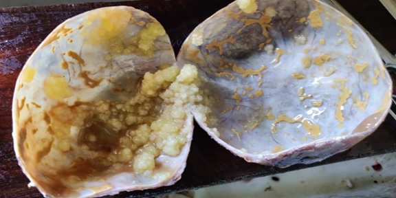
Histopathological study of ovarian lesions at a tertiary rural hospital
Abstract
Background:Ovaries are the third leading site of cancer among women trailing behind cervical and breast cancer according to the Indian cancer registries. The spectrum of ovarian lesions is wide with harmless simple cystic lesions at one end and the fatal aggressive malignant lesions at the other end. Thus, ovarian neoplasm offers a good field for research. The present study is aimed to observe the frequency of neoplastic and non-neoplastic lesions in a tertiary care centre of rural India.
Methods: This was a prospective two years observational study and was conducted at the Department of Pathology SRTR GMC, Ambajogai, including 185 ovarian lesions.
Result: Total 185 ovarian lesions were studied, 101 cases were non-neoplastic while 84 cases were neoplastic. The most common non-neoplastic lesion was corpus luteal cyst (27.7%) followed by simple ovarian cyst (24.7%) and follicular cyst (21.8%). Among 84 neoplastic cases, 74 cases (88.1%) were benign,02 cases (2.4%) were borderline tumours and 08 cases (9.5%) were diagnosed malignant. Surface epithelial tumours contributed 75% cases, while germ cell tumours and sex-cord stromal tumourcontributed 20.8% and 4.8% respectively.
Conclusions: Both non-neoplastic as well as neoplastic lesions of ovary often present with similar clinical and radiological features. So histopathological study is essential to diagnose ovarian tumours and predict their prognosis.
Downloads
References
Ellenson LH, Pirog EC, Kumar V, Abbas AK, Fausto N, Aster JC (Eds). The female genital tract: Robbins and Cotran Pathologic Basis of Disease. 8th ed; Philadelphia, Elsevier Saunders. 2010:1039-1052.
Sharma I, Sarma U, Dutta UC. Pathology of ovarian tumour- A hospital-based study. Int J Med SciClin Invention. 2014;1:284-286.
Supriya B.R, Patel R, Patel M. Histopathological evaluation of Non-Neoplastic and Neoplastic Lesions of Uterine Cervix at tertiary care centre. Trop J Path Micro 2019;5(3):177-182. Available from: https://pathology.medresearch.in/index.php/jopm/article/view/245.
Scully RE, Young RH, Clement PB. Tumors of the ovary, maldeveloped gonads, fallopian tube, and broad ligament. Amer Reg Pathol. 1998.
Selvaggi SM. Tumors of the ovary, maldeveloped gonads, fallopian tube, and broad ligament. Arch Pathol Lab Med. 2000;124(3):477.
Murthy NS, Shalini S, Suman G, Pruthvish S, Mathew A. Changing trends in incidence of ovarian cancer – The Indian scenario. Asian Pac J Cancer Prev. 2009;10(6):1025-1030.
Patel A, Patel NV. Study of frequency and histopathological pattern of soft tissue tumours in tertiary care centre of Gandhinagar, Gujarat. Trop J PatholMicrobiol. 2019;5(1):20-25. Available from: https://pathology.medresearch.in/index.php/jopm/article/view/219.
Pradhan A, Sinha AK, Upreti D. Histopathological patterns of ovarian tumors at BPKIHS. Health Renaissance. 2012;10(2):87-97. doi: https://doi.org/10.3126/hren.v10i2.6570.
Kumar P, Malhotra N. Jeffcoate’s principles of gynaecology. Jaypee Brothers Medical Publishers (P) Ltd.7th ed: 2008.
Chae MS, Kim JW, Jung JY, Choi HJ, Chung HS, Park CS, et al. CT and MR imaging of ovarian tumours with emphasis on differential diagnosis. Radiograph. 2002;22(6):1305-1325. doi: https://doi.org/10.1148/rg.226025033.
Ness RB, Grisso JA, Cottreau C, et al. Factors related to inflammation of the ovarian epithelium and risk of ovarian cancer. Epidemiol. 2000;11(2):111-117. doi: https://doi.org/10.1097/00001648-200003000-00006.
Zhang J, Ugnat AM, Clarke K, Mao Y. Ovarian cancer histology specific incidence trends in Canada 1969-1993: age-period-cohort analyses. Br J Cancer. 1999;81(1):152-158. doi: https://doi.org/10.1038/sj.bjc.6690665.
Prat J, D'Angelo E, Espinosa I. Ovarian carcinomas: at least five different diseases with distinct histological features and molecular genetics. Human Pathol. 2018;80:11-27.doi: https://doi.org/10.1016/j.humpath.2018.06.018.
Prat J. Pathology of the ovary. Philadelphia: WB Saunders Company. 2004; p. 223–7.
Jensen K. Theory and practice of histological techniques.
Bancroft JD, Gamble M, editors. Theory and practice of histological techniques. Elsevier Health Sci. 2008.
Parvatala A, Prasad JR, Rao NB, Ghanta S. Study of Non-Neoplastic Lesions of the Ovary. IOSR J Dent Med Sci. 2015;14(1):92-96. doi: https://doi.org/10.9790/0853-14168791.
Kanthikar SN, Dravid NV, Deore PN, Nikumbh DB, SuryawanshiKH.Clinico- histopathological analysis of neoplastic and non-neoplastic lesions of the ovary: a 3-year prospective study in Dhule, North Maharashtra, India. J ClinDiagn Res: JCDR. 2014;8(8):FC04–FC07. doi: https://doi.org/10.7860/JCDR/2014/8911.4709.
Makwana H, Maru A, Lakum N, Agnihotri A, Trivedi N, Joshi J. The relative frequency and histopathological pattern of ovarian masses – 11-year study at tertiary care centre.Int J Med Sci Public Health. 2014;3(1):81-84. doi: https://doi.org/10.5455/ijmsph.2013.061020132.
Singh M, Jha KK, Kafle SU, Rana R, Gautam P. Histopathological Analysis of Neoplastic and Non-Neoplastic Lesions of Ovary: A 4 Year Study in Eastern Nepal. Birat J Health Sci. 2017;2(2):168-174. doi: https://doi.org/10.3126/bjhs.v2i2.18519.
Sawant A, Mahajan S. Histopathological study of ovarian lesions at a tertiary health care institute. MVP J Med Sci. 2017;4(1):26-9. doi: http://dx.doi.org/10.18311/mvpjms/0/v0/i0/724.
Zaman S, Majid S, Hussain M, Chughtai O, Mahboob J, Chughtai S. A retrospective study of ovarian tumours and tumour-like lesions. J Ayub Medical College Abbottabad. 2010;22(1):104-108.
ModePalli N, Venugopal SB. Clinicopathological study of surface Epithelial tumours of the Ovary: An institutional study. J Clin DiagnosRes: JCDR. 2016;10(10):01-04. doi: http://dx.doi.org/10.7860/JCDR/2016/21741.8716.
Chandekar SA, Deshpande SA, Muley PS. A Clinico-Pathological Study of 120 Cases of Ovarian Tumors in a Tertiary Care Hospital. Int J Contemp Med Res. 2018;5(5):9-13. doi: http://dx.doi.org/10.21276/ijcmr.2018.5.5.35.
Sudha V, Harikrishnan V, Sridevi M, Priya P. Clinicopathological correlation of ovarian tumors in a tertiary care hospital. Indian J Pathol Oncol. 2018;5(2):332-337. doi: http://dx.doi.org/10.18231/2394-6792.2018.0062.
Prakash A, Chinthakindi S, Duraiswami R, Indira.V. Histopathological study of ovarian lesions in a tertiary care center in Hyderabad, India-a retrospective five-year study. Int J Adv Med. 2017;4(3):745-749. doi:http://dx.doi.org/10.18203/2349-3933.ijam20172265.
Jha R, Karki S. Histological pattern of ovarian tumors and their age distribution. Nepal Med Coll J. 2008;10(2):81-85.

Copyright (c) 2020 Author (s). Published by Siddharth Health Research and Social Welfare Society

This work is licensed under a Creative Commons Attribution 4.0 International License.


 OAI - Open Archives Initiative


