
A comparative study of Pap smear cytology and histopathology of cervix biopsy
Abstract
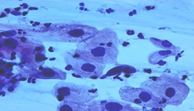
Background: Carcinoma cervix is one of the leading causes of death of the female population in developing countries. By virtue of its accessibility, cancer of the cervix can be readily diagnosed even in its preinvasive stage. If treated in the earlier stages the patient can often be cured of the disease. Carcinoma of the cervix is the fourth most frequent cancer in women worldwide and the leading cause of death from cancer in several developing countries including India. The use of cervical smear (Papanicolaou/Pap) as a screening tool has significantly reduced the incidence of cervical cancer. The present study has been carried out to evaluate the pattern of cervical cytology and its correlation with histopathological findings.
Materials and Methods: The present study was carried out in the department of pathology in a tertiary care hospital over a period of two years. In the two year study period, 2568 pap smears were received. Among these, 194 abnormal smears were identified and they were categorized under The Bethesda System 2014. The histopathological examination of biopsy cervix results of the smears was compared and analyzed.
Results: Out of 194 smears studied, reactive changes was 69 (35.57%). Atypical squamous cells of undetermined significance were 47 (24.23%). Low grade squamous intraepithelial lesions were 3 (1.55%) and high grade squamous intraepithelial lesions were 29 (14.95%). Atypical endocervical cells were 2 (1.03%). Atypical endocervical cells favor neoplastic was 4 (2.06%) and endocervical adenocarcinoma in situ was 1 (0.5%). Squamous cell carcinoma was 39 (20.10%). The histopathological examination of biopsy cervix results of the above smears was compared and analyzed. In the histopathological examination results, non-neoplastic lesions were 90 (46.4%), premalignant lesions were 31(16%) and malignant lesions were 73 (37.6%).
Conclusion: This correlative study of Pap smear and histopathological examination of the cervix revealed the overall sensitivity of 97%, the specificity of 74% and an accuracy of 87%. The false-negative and false-positive cases in this study can be minimized by proper sampling, screening, interpretation and further follow up study of repeat smears.
Downloads
References
Bray F, Ferlay J, Soerjomataram I, Siegel RL, Torre LA, Jemal A. Global cancer statistics 2018: GLOBOCAN estimates of incidence and mortality worldwide for 36 cancers in 185 countries. CA Cancer J Clin. 2018;68(6):394-424. doi: https://doi.org/10.3322/caac.21492.
Drew PA, Wilkinson EJ. Conventional cytology In: Apgar BS, Brotzman GL, Spitzer M, editors. Colposcopy principles and practice, an integrated textbook and Atlas. 2nd Ed. Philadelphia PA: Saunders Elsevier; 2008. p. 59-55.
Arbyn M, Herbert A, Schenck U, Nieminen P, Jordan J, McGoogan E, et al. European guidelines for quality assurance in cervical cancer screening: recommendations for collecting samples for conventional and liquid‐based cytology. Cytopathol. 2007;18(3):133-139. doi: https://doi.org/10.1111/j.1365-2303.2007.00464.x.
Ferlay J, Soerjomataram I, Ervik M, Forman D, Bray F, Dixit R, et al. GLOBOCAN 2012, Cancer Incidence and Mortality Worldwide in 2012. Lyon, France: International Agency for Research on Cancer; 2012.
Institute for Health Metrics and Evaluation. The Challenge Ahead: Progress in Breast and Cervical Cancer. Institute of Health Metrics and Evaluation. 2011.
Ellenson LH, Pirog EC. The female genital tract In: Kumar V, Abbas AK, Fausto N, Aster JC. Robbins and Cotran Pathological basis of disease 8th Edition Philadelphia: Saunders Elsevier 2012; 1005-1064.
Morrison AS. Screening in Chronic Disease. 2nd ed. New York: Oxford University Press; 1992. Introduction; pp. 3–42.
Welch HG, Black WC. Evaluating randomized trials of screening. J Gen Intern Med. 1997;12(2):118-124. doi: https://dx.doi.org/10.1046%2Fj.1525-1497.1997.00017.x.
Wiener HG, Klinkhamer P, Schenck U, Arbyn M, Bulten J, Bergeron C, Herbert A. European guidelines for quality assurance in cervical cancer screening: recommendations for cytology laboratories. Cytopathol. 2007; 18(2):67-78. doi: https://doi.org/10.1111/j.1365-2303.2007.00451.x.
Saslow D, Runowicz CD, Solomon D, Moscicki AB, Smith RA, Eyre HJ, et al. American Cancer Society Guidelines for the early detection of Cervical neoplasia and cancer. CA Cancer J Clin. 2002;52(6):342-362. doi: https://doi.org/10.3322/canjclin.52.6.342.
Sawaya GF, Brown AD, Washington AE, Garber AM. Clinical practice: current approaches to cervical cancer screening. N Engl J Med. 2001;344(21):1603-1607. doi: https://doi.org/10.1056/nejm200105243442107.
Bal S M, Goyal R. Detection of abnormal cytology in PAP smear. Cyto J. 2012;29(l).45-47. Available from: http://www.jcytol.org/text.asp?2012/29/1/45/93222.
Banik U, Bhattacharjee M: Pattem of epithelial cell abnormality in Pap smear: A clinicopathological and demographic correlation. Cyto J. 2011;8(8). doi: https://doi.org/10.4103/1742-6413.80527.
Sohail R, Nazir R: Evaluaion of pap smear in women attending the gynaecological opd. J Surg Pak. 2008;13(3):121-123.
Ghazal S: Pattern and factors affecting Pap smear test in Nablus, a retrospective study. Middle East J Family Med. 2004;4(4).
Sherwani R.K., Khan T., Akhtar K., Zeba A., Siddiqui FA, Rahman K., Afsan N. Conventional Pap Smear and Liquid Based Cytology for Cervical Cancer Screening- A Comparative Study. J Cytol. 2007;24(4):167-172. Available from: http://www.jcytol.org/text.asp?2007/24/4/167/41888.
Bukhari H, Saba K: Clinicopathological importance of Papanicolaou smears for the diagnosis of premalignant and malignant lesions of the cervix. J Cytol 2012;29(1):20-25. Available from: http://www.jcytol.org/text.asp?2012/29/1/20/93213.
Chhabra Y, Behera BG, Khalkho J, Pati N. Cytomorphological study of PAP smears in precancerous and cancerous lesions. J Cytol. 2003;20(2):64-67.
Ozkara SK, Yıldız K. Retrospective five year analysis of our servicovaginal cytology screening programme under the perspective of Bethesda-2001. Turk Bullet Pathol. 2002; 19:119-124.
Yeah GP, Chan KW. The Accuracy of Papanicoaou smear predictions: cytohistological correlation of 283 cases. Hong Kong Med J. 1997;3:373-376.
Gupta S, Sodhani P. Why is high grade squamous intraepithelial neoplasia under-diagnosed on cytology in a quarter of cases? Analysis of smear characteristics in discrepant cases. Indian J Cancer. 2004;41(3):104-108.
Anderson MB. Jones BA. False positive cervico vaginal Cytology. A follow up study – Actacytol. 1997;41(6):1697-1700. doi: https://doi.org/10.1159/000333170.
Tritz: Discrepancies between cytologic and histologic diagnoses, Koss diagnostic cytology and its histopathologic bases, 5th edition, Lippincot Williams, 2006, Vol – 1: 364-368.
Mitch, Medley: Correlation between cytology and biopsy cervix, Koss diagnostic cytology and its histopathologic bases 5th edition, Lippincot Williams, 2006, Vol – 1: 364-368.
Boyes DA, Nichols AM, Millner AM, Worth AJ. Recent results from the British Columbia screening program for cervical cancer. Amer J Obstet Gynaecol. 1977;128(6):692-693. doi: https://doi.org/10.1016/0002-9378(77)90223-X.
Dhakal R, Makaju R, Sharma S, Bhandari S, Shreshtha S, Bastakoti R. Correlation of Cervical Pap Smear with Biopsy in the Lesion of Cervix. Kathmandu Univ Med J. 2016;55(3):254-257.
Atla BL, Uma P, Shamili M, Kumar SS. Cytological patterns of cervical pap smears with histopathological correlation. Int J Res Med Sci. 2015;3(8):1911-1916. doi: http://dx.doi.org/10.18203/2320-6012.ijrms20150300.
Copyright (c) 2020 Author (s). Published by Siddharth Health Research and Social Welfare Society

This work is licensed under a Creative Commons Attribution 4.0 International License.

 OAI - Open Archives Initiative