Histopathological evaluation of Non-Neoplastic and Neoplastic Lesions of Uterine Cervix at tertiary care centre
Abstract
Background: Uterine cervix is common specimen from gynecological department with non-neoplastic and neoplastic lesions. Most cervical cancers can be detected at pre-invasive state with an adequate screening and treated appropriately thus preventing overt progression to full blown cancer and hence decreasing morbidity and mortality. Carcinoma of cervix is a preventable tumor and if good effort given in detecting preinvasive lesion one can give definite treatment at earliest.
Objectives: To evaluate the histopathological diagnosis of a biopsy of cervix in women with unhealthy cervix. Also, to study the agerelated, incidence and the incidence of various nonneoplastic and neoplastic lesions of the cervix.
Methodology: It is a cross sectional study. Institutional Ethics committee permission was taken prior to start of the study. Pertinent clinical history like age, significant family and personal history, history of other diseases was taken. Cervical biopsy specimens collected from histopathology section and grading of cervical lesions was done based on the proportion of stained cells and the intensity of staining. Statistical analysis was done. The data were tabulated and frequencies and percentages were calculated.
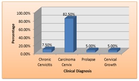
Observations: The common age group was 41-50 years (37.50%) followed by 31-40 years of age (26.25%). The symptoms with which patients presented were vaginal bleeding (52.50%) followed by white discharge per vagina (16.25%) and menorrhagia (12.50%). The premalignant lesions were seen in 12 cases (15.0%) and malignant lesions in 68 cases (85.0%). Among them 64 malignant tumours were epithelial in origin. As per microscopic examinations squamous cell carcinoma was diagnosed in 61 cases (93.75%), followed by 2 cases (3.13%) of adenocarcinoma, and 1 case (1.58%) of adenosquamous carcinoma and 1 case (1.54%) of rhabdomyosarcoma.
Conclusion: Cervical cancer continues to be the most common cancer of females in developing countries. One of the most significant advances in the management of cervical neoplasms has been the realization that cervical intraepithelial lesions behave as progressive stages of a biologic continuum towards the development of invasive cancer.
Downloads
References
2. Anderson MC. The etiology of cervical cancer. Female Reproductive System in Systemic Pathology. 3rd ed., Vol 6 New York: Churchill Livingstone; 1991:76-9.
3. Wells M, Ostor AG, Crum CP, Franceschi S, Tommasino M, Nesland JM, et al. Epithelial tumours. In: Tavassoli FA, Devilee P, editors. World Health Organisation classification of tumours. Pathology and genetics of tumours of the breast and female genital organs. Lyon: IARC Press-2003.p.262-79.
4. Akhter S, Bari A, Hayat Z. Variability study between Pap smear, Colposcopy and Cervical Histopathology findings. J Pak Med Assoc. 2015;65(12):1295-9.[pubmed]
5. Mehta A, Ladola H, Kotadiya K, Edwin R, Patel V, Patel V. Study of high risk cases for early detection of cervical cancer by PAP’s smear and visual inspection by Lugol’s Iodine Method. NHL J Medical Sciences. 2013;2(1):65-8.
6. Atla BL, Uma P, Shamili M, Kumar S. Cytological patterns of cervical pap smears with histopathological correlation. Int J Res Med Sci. 2015;3(8):1911-6.
7. Sharma A, Rajappa M, Saxena A, Shamla M. Telomerase activity as a tumour marker in Indian women with cervical intraepithelial neoplasia and cervical cancer. Mol DiagTher 2007;11(3):193-201.[pubmed]
8. Sherman ME, Wang SS, Tarone R, et al. Histopathologic extent of cervical intraepithelial neoplasia 3 lesions in the atypical squamous cells of undetermined significance low-grade squamous intraepithelial lesion triage study: implications for subject safety and lead-time bias. Cancer Epidemiol Biomarkers Prev. 2003 Apr;12(4):372-9.[pubmed]
9. Solapurkar ML. Histopathology of uterine cervix in malignant and benign lesions. J ObstetGynaecol India 1985;35:933-7.
10. Momtahen S, Kadivar M, Kazzazi AS, et al. Assessment of gynecologic malignancies: a multi-center study in Tehran (1995-2005). Indian J Cancer. 2009 Jul-Sep;46(3):226-30. doi: 10.4103/0019-509X.52957.
11. Chhabra S, Bhavani M, Mahajan N, et al. Cervical cancer in Indian rural women: trends over two decades. J ObstetGynaecol. 2010;30(7):725-8. doi: 10.3109/01443615.2010.501412.[pubmed]
12. Lowe D, Jorizzo J, Chiphangwi J, Hutt MS. Cervical carcinoma in Malawi: a histopathologic study of 260 cases. Cancer. 1981 May 15;47(10):2493-5.[pubmed]
13. Scarinci IC1, Garcia FA, Kobetz E, et al. Cervical cancer prevention: new tools and old barriers. Cancer. 2010 Jun 1;116(11):2531-42. doi: 10.1002/cncr.25065.[pubmed]
14. Chen J, Macdonald K, Gaffney DK. Incidence, mortality, and prognostic factors of small cell carcinoma of the cervix. ObstetGynecol 2008; 111: 13 94-402.[pubmed]
15. Maiman MA, Fruchter RG, DiMaio TM, et al. Superficially invasive squamous cell carcinoma of the cervix. Obstet Gynecol. 1988 Sep;72(3 Pt 1):399-403.[pubmed]
16. van Nagell JR Jr, Greenwell N, Powell DF, et al. Microinvasive carcinoma of the cervix. Am J Obstet Gynecol. 1983 Apr 15;145(8):981-91.[pubmed]
17. Hurt WG, Silverberg SG, Frable WJ, et al. Adenocarcinoma of the cervix: histopathologic and clinical features. Am J Obstet Gynecol. 1977 Oct 1;129(3):304-15.[pubmed]
18. Zanetta GM, Gostout BS, Kerney GL, Lee R.A. An unusual presentation of cervical adeno-squamous carcinoma. J Pelvic Surg 1999;5(3):171-4.


 OAI - Open Archives Initiative


