
Study of frequency and histopathological pattern of soft tissue tumours in tertiary care centre of Gandhinagar, Gujarat
Abstract
Background: Ovaries are complex intra-pelvic organs of the female reproductive system. Ovarian cancer accounts for 3% of all cancers in females and is the fifth most common cause of cancer death in women. Early menarche, late menopause, nulliparity and high socioeconomic status are associated with an increased risk for ovarian neoplasms. Histo-pathological diagnosis remains the mainstay to differentiate neoplastic lesions from non-neoplastic lesions.
Aims and objectives: This study aims to analyze the view of ovarian tumors with respect to clinical presentation, gross and microscopic characteristics and also to study the frequency and histopathological patterns of ovarian tumours.
Materials and Methods: This study comprised of 100 cases of ovarian tumours received in the Department of Pathology, GMERS Medical College, Gandhinagar were analysed. Their gross features, microscopic findings were analysed in detail. Ovarian tumours were divided into benign and malignant categories and their further sub typing were done according to WHO Classification.
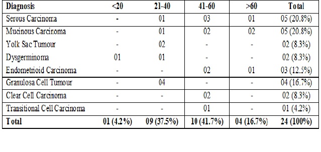
Results: Out of total 100 cases, 76 were benign and 24 were malignant. Out of 100 cases, 62% were between 21-40 years of age followed by 28% were between 41-60 years of age. Most common presenting symptom was pain in abdomen followed by lump in abdomen and heavy menstruation. Out of total 100 cases, 70% were surface epithelial tumours, 24% were germ cell tumours and 6% were sex cord stromal tumours.
Conclusion: To conclude we recommend microscopic histopathological examination of every ovarian mass in order to assess the importance of pathological grading and staging and they must be classified correctly so that the patient can be provided with appropriate treatment and prognosis.
Downloads
References
2. Birare SD, Dale AR. Clinicopathological study of ovarian tumours: a 5 year study. Ind J Pathol Oncol, 2018;5(3):360-365. DOI: 10.18231/2394-6792.2018.0070
3. Montag A, Kumar V. The female genital system and breast. Robbins Basic Pathology. 9th Ed. Philadelphia: Saunders Elsevier. 2014:991-1042.
4. Mankar DV, Jain GK. Histopathological Profile of Ovarian tumours. A twelve year experience. Muller J Med Sci Res. 2015;6(2):107-11. DOI: 10.4103/0975-9727.16067
5. Murthy NS, Shalini S, Suman G, et al. Changing trends in incidence of ovarian cancer - the Indian scenario. Asian Pac J Cancer Prev. 2009;10(6):1025-30.[pubmed]
6. Kurman RJ, Ellenson LH, Ronnett BM, Blaustein’s pathology of female genital tract, 6th Ed., New York: Springer:2011.p.680-2.
7. JaunRosai, “Rosai and Ackerman’s Surgical Pathology,” 9th Ed., New Delhi, Elsevier 2004.
8. Aziz S, Kuperstein G, Rosen B, et al. A genetic epidemiological study of carcinoma of the fallopian tube. Gynecol Oncol. 2001 Mar;80(3):341-5.[pubmed]
9. Narod SA, Boyd J. Current understanding of the epidemiology and clinical implications of BRCA1 and BRCA2 mutations for ovarian cancer. CurrOpinObstet Gynecol. 2002 Feb;14(1):19-26.[pubmed]
10. Novak ER, and Woodruff JD. In:Novak’sGyneacologic and Obstetric pathology.
11. Rosai J. Rosai and Ackerman’s Surgical Pathology. Tenth Ed., New York: Mosby Elsevior;2011.p.1562-3.
12. Sharma I, Sharma U, Dutta UC. Pathology of Ovarian Tumours – A Hospital based study. International Journal of Medical Science and Clinical Invention. 2014;1(6):284-6.
13. Ellenson LH, Pirog EC. Robbins and Cotran pathologic basis of disease. 9th Ed. Philadelphia (USA), Elsevior and Saunders Press; 2014. Chapter 22, The Female Genital Tract; p.991-1042.
14. Gilks CB, Prat J. Ovarian carcinoma pathology and genetics: recent advances. Hum Pathol. 2009 Sep;40(9):1213-23. doi: 10.1016/j.humpath.2009.04.017. Epub 2009 Jun 24.[pubmed]
15. S.N.Kanthikar. “Clinico-Histopathological Analysis of Neoplastic and Non-Neoplastic Lesions of the Ovary: A year Prospective Study in Dhule, North Maharashtra, India. J Clin Diagn Res. 2014;8(8):04-07. DOI: 10.7860/JCDR/2014/8911.4709
16. Mondal SK1, Banyopadhyay R, Nag DR, et al. Histologic pattern, bilaterality and clinical evaluation of 957 ovarian neoplasms: a 10-year study in a tertiary hospital of eastern India. J Cancer Res Ther. 2011 Oct-Dec;7(4):433-7. doi: 10.4103/0973-1482.92011.
17. Robert J. Kurman, Maria Luisa Carcangiu, C. Simon Herrington, Robert H. Young, (Eds.): WHO Classification of Tumours of Female Reproductive Organs 4th Edition IARC Lyon. 2014.p.12-13.
18. Abdullah LS1, Bondagji NS. et al. Histopathological pattern of ovarian neoplasms and their age distribution in the western region of Saudi Arabia. Saudi Med J. 2012 Jan;33(1):61-5.
19. Neethu GV, Divya P, Preethi CR, Rajashekar KS, Soumya BM. Histopathological study of ovarian tumours. Ind J Pathol Oncol, 2018;5(1):25-8. DOI:10.1823/2394-6792.2018.0005.
20. Jha R, Karki S. Histological pattern of ovarian tumors and their age distribution. Nepal Med Coll J. 2008 Jun;10(2):81-5.[pubmed]
21. Ranjana Hawaldar, SadhnaSodani, Ekta Patidar. Histopathological spectrum of ovarian tumours – A two years retrospective study. Ind J Pathol Oncol, 2017;4(3):450-453. DOI: 10.18231/2394-6792.2017.0097.
22. Makwana HH, Maru AM, Lakum NR, Agnihotri AS, Trivedi NJ, Joshi JR. The relative frequency and histopathological pattern of ovarian masses – 11 year study at tertiary care centre. Ind J Med Sci Public Health, 2014;3(1):81-4. DOI: 10.5455/ijmsph.2013.061020132.
23. Gupta N, Bisht D, Agarwal AK, et al. Retrospective and prospective study of ovarian tumours and tumour-like lesions. Indian J PatholMicrobiol. 2007 Jul;50(3):525-7.[pubmed]
24. Maheshwari V, Tyagi SP, Saxena K, et al. Surface epithelial tumours of the ovary. Indian J PatholMicrobiol. 1994 Jan;37(1):75-85.[pubmed]
25. Yasmeen S, Yasmeen A. Frequency of benign and malignant ovarian tumours in a tertiary care hospital. J Postgrad Med Inst 2006;20(4):393-7.
26. Ashraf A, Shaikh AS, Ishfaq A, Akram A, Kamal F, Ahmed N. The relative frequency and histopathological patterns of ovarian masses. Biomed 2012;28(1):98-102.
27. Sheikh S, Bashir H, Farooq S, Beigh A, Manzoor F, Reshi R. Histopathological spectrum of ovarian tumours from a referral hospital in Kashmir valley, Jammu and Kashmir, India. Intl J Res Med Sci, 2017;5(5):2110-4. DOI: 10.18203/2320-6012.ijrms20171852.
28. Dhakal R, Makaju R, Bastakoti R. Clinicomorphological spectrum of ovarian cystic lesions. Kathmandu Univ Med J. 2016;14(53):13-6. PMID: 27892434.
29. Sawant A, Mahajan S. Histopathological study of ovarian lesions at a tertiary health care institute. MVP J Med Sco, 2017;4(1):26-9. DOI: 10.18311/mvpjms/0/v0/i0/724.
30. Bodal VK, Jindal T, Bal MS, Bhaga R, Kaur S, Mall N, Goyal P. A clinicopathological study of ovarian lesions. RRJMHS 2014;3(1):50-6.

 OAI - Open Archives Initiative