
Bacteriological profile of diabetic foot ulcer using hicrome UTI Agar
Abstract
Introduction: Foot ulcers are one of the common complications of diabetes mellitus. Diabetic foot ulcer infections are usually polymicrobial infections. For effective treatment, quick isolation and identification of causative organisms with appropriate antibiotic susceptibility testing is needed.
Objective: Isolation and identification of bacteria using routine media and HiCrome UTI agar.
Materials and Methods: Samples were taken from all Type II diabetes mellitus patients with foot ulcers attending hospital. Samples were collected from deeper portion of the ulcer using 2 sterile swabs and processed using conventional methods and HiCrome UTI agar. Antibiotic susceptibility testing was done on Mueller Hinton agar according to CLSI guidelines.
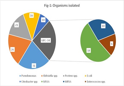
Results: Among 100 samples tested, 138 organisms were isolated as 38% of samples yielded mixed growth. Conventional methods failed to detect 6 (4.3%) isolates form mixed cultures. HiCrome UTI agar isolated all organisms in the cultures including 4 isolates of enterococci and 2 isolates of MSSA, which were missed in conventional methods (p < 0.01). Pseudomonas (22.4%) was commonest organism isolated. Followed by Klebsiella spp. (18.8%), Proteus spp. (15.2%), MSSA (13%), Escherichia coli (11.5%), Citrobacter spp. (7.2%), MRSA (7.2%), Enterococcus spp. (4.3%).
Conclusion: Gram negative organisms (75.3%) were predominantly isolated in the study. HiCrome UTI agar can be used for primary identification and quick isolation of organisms where facilities for routine culturing are not available. It is both sensitive and specific in isolating and identifying organisms as in polymicrobial infections.
Downloads
References
2. Mehta VJ, Kikani KM, Mehta SJ. Microbiological profile of diabetic foot ulcers and its antibiotic susceptibility pattern in a teaching hospital, Gujarat. Int J Basic Clin Pharmacol. (2014), February 15, 2016; 3(1): 92-95. DOI: 10.5455/2319-2003.ijbcp.20140209.
3. Yerat RC, Rangasamy VR. A clinicomicrobial study of diabetic foot ulcer infections in South India. Int J Med Public Health 2015; 5: 236-41. DOI: 10.4103/2230-8598.161545.
4. Sugandhi P, Prasanth D A. Bacteriological Profile of Diabetic Foot Infections. IJIRSET. 2014; Vol. 3, Issue 7, 14688-14692.
5. Yazdanpanah L, Nasiri M, Adarvishi S. Literature review on the management of diabetic foot ulcer. World J Diabetes. 2015 Feb 15;6(1):37-53. doi: 10.4239/wjd.v6.i1.37. [PubMed]
6. Jayashri P, Payal R D, Sanjay R, Afroz B, Bimal C, Parul D S. Utility of UTI CHROM agar media for the rapid identification of uropathogens. NHL journal of medical sciences. 2013; Vol 2 (1): 39-42.
7. Zubair M, Malik A, Ahmad J. Diabetic Foot Ulcer: A Review. American Journal of Internal Medicine. Vol. 3, No. 2, 2015, pp. 28-49. DOI: 10.1016/j.dsx.2015.04.007.
8. Clinical And Laboratory Standards Institute. Performance Standards For Antimicrobial Susceptibility Testing.M100-S24, Vol 34, no 1, Wayne, Pennsylvania; 2016, p.38-42.
9. Tiwari S, Pratyush D, Dwivedi A, Gupta S, Rai M, Singh S (2012) Microbiological and clinical characteristics of diabetic foot infections in northern India. The Journal Of Infection In Developing Countries 6 (04): 329-332. DOI: https://doi.org/10.3855/jidc.1827.
10. Dwedar R, Ismail D K and Abdulbaky A. Diabetic foot Infection: Microbiological Causes with Special Reference to their Antibiotic Resistance Pattern. Egyptian Journal of Medical Microbiology Volume 24 / No. 3 / July 2015 95-102.
11. Chaudhry W. N., Badar, R., Jamal M., Jeong J., Zafar J., Andleeb S. Clinico microbiological study and antibiotic resistance profile of mecA and ESBL gene prevalence in patients with diabetic foot infections. Experimental and Therapeutic Medicine 11.3 (2016): 1031-1038. DOI: 10.3892/etm.2016.2996.
12. Patil SV, Mane RR. Bacterial and clinical profile of diabetic foot ulcer using optimal culture techniques. Int J Res Med Sci 2017;5: 496-502. DOI: http://dx.doi.org/10.18203/2320-6012.ijrms20170139.

 OAI - Open Archives Initiative