
Histopathogical study of dermatological lesions in HIV reactive patients
Abstract
Background: Skin lesions are commonly encountered in HIV patients (>90%). Several skin diseases have proved to be sensitive and useful indicators of progression of HIV infection. Although these conditions may be seen in general healthy population, their occurrence in patients with acquired immunodeficiency syndrome is often atypical, more severe and explosive.
Objective: The present study was carried out to categorize the prevalence of different skin lesions in HIV positive patients, their clinical and histopathological analysis and role of skin biopsy in their diagnosis.
Materials and Methods: A total 60 known HIV positive patients with the symptomatic skin lesions were included and their histopathological and clinical correlation and biopsy were analyzed.
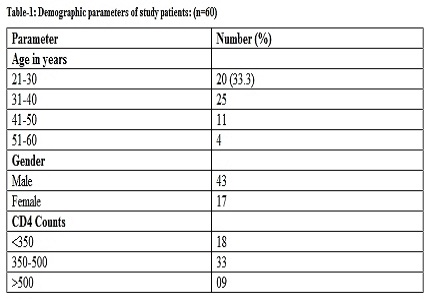
Results: Maximum patients were between 31 and 40 years (41.6 %). Papular lesions and dermatitis are the most common lesions seen in patients having CD4 count > 350 and viral and fungal infections are most common lesions seen in patients having CD4 count < 350. Out of 60 cases, 10 (16.6%) show features of papular lesions (including Pruritic Papular eruptions, Psoriasis, Seborrhoic dermatitis, Scaly lesion, Eosinophilic folliculitis, Lichen planus), 8(13.3%) show features of dermatitis (including Atopic Dermatitis, Ashy Dermatitis, Urticaria, hyperpigmented patch, Chronic Non-specific dermatitis), and 8 (13.3%) show features of bacterial infections (including Chanchroid, Hansen, TBVC, Folliculitis, pustular lesion, lupus vulgaris, syphilis, pyoderma gangrenosum). Histopathological examination showed high sensitivity of 88.8% and specificity of 96.0% in diagnosis of papular lesions.
Conclusions: Morphological pattern of skin lesions in HIV is often nondiagnostic. Histopathological correlation is therefore pivotal in the accurate diagnosis of many HIV induced skin diseases. The histopathological examination of such lesions helps in confirming the diagnosis as they have high sensitivity and specificity values.
Downloads
References
2. UNAIDS. Report on global AIDS epidemic July; 2004.
3. Park AG. Text book of preventive and social medicine. 7th ed. JP publication; 2007.
4. Grayson W. The HIV-positive skin biopsy. J Clin Pathol. 2008 Jul;61(7):802-17. Epub 2007 Nov 15. [PubMed]
5. Coldiron BM, Bergstresser PR. Prevalence and clinical spectrum of skin disease in patients infected with human immunodeficiency virus. Archives of dermatology. 1989 Mar 1;125(3):357-61. [PubMed]
6. Tschachler E, Bergstresser PR, Stingl G. HIV-related skin diseases. Lancet. 1996 Sep 7;348(9028):659-63. [PubMed]
7. Jindal N, Aggarwal A, Kaur S. HIV seroprevalence and HIV associated dermatoses among patients presenting with skin and mucocutaneous disorders. Indian Journal of Dermatology, Venereology, and Leprology. 2009 May 1;75(3):283.
8. Kumarasamy N, Vallabhaneni S, Flanigan TP, Mayer KH, Solomon S. Clinical profile of HIV in India. Indian J Med Res. 2005 Apr;121(4):377-94. [PubMed]
9. Wiwanitkit V. Prevalence of dermatological disorders in Thai HIV infected patients correlated with different CD4 lymphocyte count statuses: A note on 120 cases. Int J Dermatol. 2004;43:265–8.
10. Spira R, Mignard M, Doutre MS, Morlat P, Dabis F. Prevalence of cutaneous disorders in a population of HIV-infected patients: southwestern France, 1996. Arch Dermatol. 1998 Oct 1;134(10):1208-12.
11. Rad F, Ghadheri E. Relationship between skin manifestations and CD4 count among HIV positive patients. Pakistani Journal of medical sciences.2008;24:114-117.
12. NACO. National AIDS Control Organization Annual Report. New Delhi, India; 2007.
13. Supanaranond W, Desakorn V, Sitakalin C, Naing N, Chirachankul P. Cutaneous manifestations in HIV positive patients. Southeast Asian J Trop Med Public Health. 2001 Mar;32(1):171-6. [PubMed]
14. Hevia O, Jimenez-Acosta F, Ceballos PI, Gould EW, Penneys NS. Pruritic papular eruption of the acquired immunodeficiency syndrome: a clinicopathologic study. J Am Acad Dermatol. 1991 Feb;24(2 Pt 1):231-5.
15. Rosenthal D, LeBoit PE, Klumpp L, Berger TG. Human immunodeficiency virus-associated eosinophilic folliculitis. A unique dermatosis associated with advanced human immunodeficiency virus infection. Arch Dermatol. 1991 Feb;127(2):206-9.
16. Nichols L, Balogh K, Silverman M. Bacterial infections in the acquired immune deficiency syndrome. Clinicopathologic correlations in a series of autopsy cases. Am J Clin Pathol. 1989;92:787–90.
17. Dover JS, Johnson RA. Cutaneous manifestations of human immunodeficiency virus infection. Part II. Arch Dermatol. 1991 Oct;127(10):1549-58.
18. Vasudevan B, Sagar A, Bahal A, Brig AP, Mohanty VS. Cutaneous manifestations of HIV – A detailed study of morphological variants, markers of advanced disease, and the changing spectrum. Med J Armed Forces India. 2012;68:20–7.

 OAI - Open Archives Initiative