
Cytology- A useful diagnostic tool in ascites, 2 years study
Abstract
Background: Identification of malignant cells in any body fluid always is a challenging task for cytopathologist. Detailed clinical history, morphological evaluation and sometimes modern techniques like cytochemistry and immunohistochemistry can help us whenever required. Ascites is a common clinical finding with a wide range of etiology. Ascites is defined as collection of excessive fluid in the peritoneal cavity. The present study aims to note down various pathological findings in Transudative and Exudative ascites. This will help the clinician, surgeon and oncologist in treating the patient and it is the determining factor for treating patient.
Materials and Methods: It is an observational, prospective study. 120 cases of ascitic fluid cytology were collected from the patients admitted in a tertiary care hospital which is situated in Ahmedabad over a period of 2 years. Detailed examination – physical, biochemical and microbiological (wherever indicated) was done along with cytology examination of ascitic fluid.
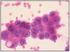
Result:We studied 120 cases of ascitic fluid cytology. Most common causes of ascitic fluid effusion after examining 120 cases were liver cirrhosis followed by tuberculosis, inflammatory, malignant, acute bacterial infection, cardiac and renal causes. Among malignant effusion GIT and ovary were most common primary site of malignancy and commonest malignancy was adenocarcinoma.
Conclusion: Definite diagnosis of ascitic fluid effusion can be done in most of the cases by doing cytology and detailed examination like physical, biochemical and microbiological examination of that fluid. However in some cases newer techniques like cytochemistry and immunohistochemistry may require for confirmation. Cytology is first line of investigation along with other radiological investigation to rule out neoplastic lesions. It is also useful in identification of non neoplastic lesions like tuberculosis, Cirrhosis of liver, Nephrotic Syndrome, congestive cardiac failure, Anaemia-Hypoproteinemia & Spontaneous bacterial peritonitis. It helps clinician in management of patients.
Downloads
References
2. Kumavat PV, Kulkarni MP, Sulhyan KR. Cytological study of Effusions. Indian Medical Gazette. 2013;August: 306-313.
3. Turnage RH. Abdominal wall, umbilicus, peritoneum, mesenteries, omentum, and retroperitoneum. Sabiston textbook of surgery. 2008:1150-1.
4. Runyon BA. Care of patients with ascites. N Engl J Med. 1994 Feb 3;330(5):337-42.[pubmed]
5. Shobha SN, Kodandaswamy CR. Utility of modified cell block technique in cases of pleural effusion suspected of malignancy. Int J Health Sci Res. 2013;3(1):33-8.
6. Zocchi L. Physiology and pathophysiology of pleural fluid turnover. Eur Respir J. 2002 Dec;20(6):1545-58.[pubmed]
7. Culling CF, Allison RT, Barr WT. Cytology technique. cellular pathology technique. 4th edn. London: Butterworth and Co.(Publishers) Ltd. 1985;336.
8. Khan FY. Ascites in the state of Qatar: aetiology and diagnostic value of ascitic fluid analysis. Singapore Med J. 2007 May;48(5):434-9.[pubmed]
9. Mahmood G, Debnath CR, Mandal AK. Evaluation of 100 cases of ascites. Mymensingh Med J. 2009 Jan;18(1):62-6.[pubmed]
10. Chung ES, Eun SJ, Song KE, Suh JS, Lee WK, Kim JS. Diagnostic value of cholesterol and triglyceride in pleural andascitic fluid. Korean Journal of Clinical Pathology. 1992 Sep 1;12(3):291-8.
11. Jain SC, Misra SM, Misra NP, et al. Diagnostic value of ascitic fluid examination. J Assoc Physicians India. 1966 Jan;14(1):59-69.[pubmed]
12. Nath K, Mital HS, Mishra SD, et al. Diagnostic value of ascitic fluid examination. J Assoc Physicians India. 1968 Dec;16(12):991-6.[pubmed]
13. Khan TH, Durrani HA, Shah SN. Evaluation of peritoneal biopsy and ascitic fluid examination in the etiological diagnosis of ascites. J Assoc Physicians India. 1976 Sep;24(9):579-86.[pubmed]
14. Malabu UH, Olubuyide IO, Shaibu ME, Olawuyi F. Ascites in Ibadan, Nigeria–usefulness of albumin gradient in its etiologic diagnosis. Biomedical Research. 2006;17:105-9.
15. Sears D, Hajdu SI. The cytologic diagnosis of malignant neoplasms in pleural and peritoneal effusions. Acta Cytol. 1987 Mar-Apr;31(2):85-97.[pubmed]
16. Cardozo PL. A critical evaluation of 3000 cytologic analyses of pleural fluid ascitic fluid and pericardial fluid. Acta Cytologica. 1966 Jan 1;10(6):455.
17. Khan N, Sherwani RK, Afroz N, Kapoor S. Cytodiagnosis of malignant effusion and determination of primary site. Journal of cytology. 2005 Jul 1;22(3):107.

 OAI - Open Archives Initiative