
Comparative study of red blood cell morphology in peripheral smear and automated cell counter
Abstract
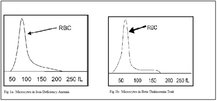
Background: Automated cell counters are a very important part of pathology laboratory for evaluation of complete blood count (CBC). They also provide RBC histograms to interpret differentmorphological variations of Red Blood Cells.These histograms and other parameters have been found veryuseful in diagnosing various hematological conditions and Red Blood Cell disorders if they are correctly interpreted.Examination of peripheral smears is still the gold standardfor diagnosing some of the RBC disorders which might not be diagnosed otherwise by automated cell counters. They play an important role in quality check of automated analyzers.
Materials and Methods:In this prospective study we have taken 200 samples over a period of 6 months. We did comparative study between RBC histograms obtained by automated hematology analyzer and peripheral blood smears stained by field stain. We have discussed morphological variations of red blood cells and their characteristic changes in respective RBC histograms.
Result:Out of 200 samples of RBC histogram interpretation, 138 cases showed correlation with peripheral smear findings while 62 cases showed discrepancies.
Conclusion: Microscopic examination of peripheral blood smear still remains gold standard for diagnosis of various hematological conditions.
Downloads
References
2. Bessman JD, Gilmer PR Jr, Gardner FH. Improved classification of anemias by MCV and RDW. Am J Clin Pathol. 1983 Sep;80(3):322-6.[pubmed]
3. Williams LJ. Cell histograms: New trends in data interpretation and cell classification. Journal of medical technology. 1984;1(3):189-97.
4. Fossat C, David M, Harle JR, Sainty D, Horschowski N, Verdot JJ, Mongin M. New parameters in erythrocyte counting. Value of histograms. Archives of pathology & laboratory medicine. 1987 Dec;111(12):1150-4.
5. Lawrence A, Young M, Cooper A, Turner E. Red cell histograms in the diagnosis of diseases. Hematology Beyond the Microscope. New York, NY: Technicon Instruments. 1984:155-64. [pubmed]
6. Beckman Coulter LH. 780 on line IB072841. Beckman Coulter Education Center, Miami Lakes, FL. 2007.
7. Lokwani DP. The ABC of CBC: Interpretation of complete blood count and histograms. JP Medical Ltd; 2013 May 30.
8. Rowan RM. Blood Cell Volume Analysis: A New Screening Technology for the Haematologist. Albert Clark;1983.
9. Steele BW, Wu NC, Whitcomb CL. White blood cell and platelet counting performance by hematology analyzers: a critical evaluation. Laboratory Hematology. 2001 Jan 1;7:255-66.
10. ART BT. High mean corpuscular hemoglobin concentration: Its causes and effects on automated CBC results. Canadian Journal of Medical Laboratory Science. 2007 May 1;69(3):113.

 OAI - Open Archives Initiative