
Cutaneous metastasis: Indicators of internal malignancy diagnosed on fine needle aspiration cytology
Abstract
Introduction: Cutaneous metastases (CM) from various malignancies are uncommon with incidence of 0.8-4% and indicate disseminated disease with poor outcome. Metastatic adenocarcinoma to the skin occurs from gastro-intestinal tract, breast, lung and ovary and can be determined by high index of suspicion.Fine needle aspiration cytology is commonly employed for its diagnosis. CM from breast carcinoma to the chest wall are not very rare; however, distant CM are far less common occurring mostly within five years of its complete resection. Gastric adenocarcinoma presenting as CM is extremely rare, with 6%males and 1%females presenting likewise. Classically, these lesions appear as slow-growing, painless, discrete hard nodules with intact overlying epidermis.
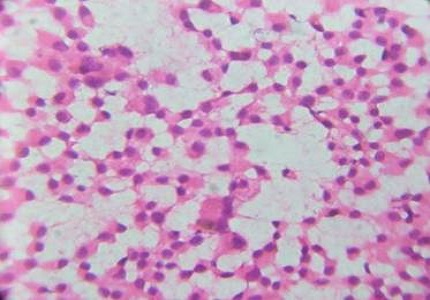
Case Report: We describe three cases of CM from malignancies of two organs & diagnosed on FNAC – 1) A 65 year male who underwent complete resection for breast carcinoma 10 years ago, presented with chest wall metastasis, 2) A 42 year female presented with distant CM following complete treatment for breast carcinoma 4 years ago, and 3) A 52 year female presented with CM nodules over abdomen after 3 years of radical gastrectomy and adjuvant chemotherapy.
Conclusion: CM can be the first sign of disseminated malignancy; primary or post-surgery. CM can also occur in response to chemotherapy. Post-surgery, such skin lesions could occur as part of dissemination from another organ malignancy (dual malignancy).FNAC is a cost-effective, precise procedure for diagnosis of CM and also helps in differentiating these from various primary cutaneous malignancies and inflammatory conditions.
Downloads
References
Brady LW, O'Neill EA, Farber SH. Unusual sites of metastases. Semin Oncol. 1977 Mar;4(1):59-64.
Rolz-Cruz G, Kim CC. Tumorinvasion of the skin. DermatolClin. 2008 Jan;26(1):89-102, viii.
Asli Turgut Erdemir, UlviyeAtilganoglu, NahideOnsun, Adnan Somay. Cutaneous Metastasis from Gastric Adenocarcinoma. Indian J Dermatol. 2011 Mar-Apr; 56(2): 236-237.
Bansal R, Patel T, Sarin J, Parikh B, Ohri A, Trivedi P. Cutaneous and subcutaneous metastases from internal malignancies: an analysis of casesdiagnosed by fine needle aspiration. Diagn Cytopathol. 2011 Dec;39(12):882-7. doi: https://doi.org/10.1002/dc.21485.
Brownstein MH, Helwig EB. Patterns of cutaneous metastasis. Arch Dermatol.1972 Jun;105(6):862-8.
Cox NH, Coulson LH. Systemic disease and the skin. In: Burns T, Breathnach S, Cox N, Griffiths C, editors. Rook’s Textbook Of Dermatology. 8th ed. West Sussex: Wiley Blackwell Publications; 2010.p.62.1-113.
Karki S, Pathak R, Manandhar U, Koirala S. Metastatic cutaneous and subcutaneous lesions: Analysis of cases diagnosed on fine needle aspiration cytology. JPN 2011;1:37-40.
Rajagopal R, Arora PN, Ramasastry CV, Kar PK. Skin changes in internal malignancy. Indian J Dermatol VenereolLeprol2004;70:221-225.
Johnson WC. Metastatic carcinoma of the skin: Incidence and dissemination. In: Elder DE, editor. Lever’s Histopathology of the Skin. 10th ed. Philadelphia: Lippincott Williams and Wilkins; 2009.p 1149-1157.
Ayyamperumal A, Tharini GK, Ravindran V, Parveen B. Cutaneous manifestations of internal malignancy. Indian J Dermatol 2012;57:260-264.
Alcaraz I, Cerroni L, Rutten A, Kutzner H, Requena L. Cutaneous Metastasis from internal malignancies: A clinicopathological and immunohistochemical review. Am J Dermatopathol2012;34:347-93.
Srinivasan R, Ray R, Nijhawan R. Metastaticcutaneous and subcutaneousdeposits from internalcarcinoma. An analysis of casesdiagnosed by fine needle aspiration. ActaCytol. 1993 Nov-Dec;37(6):894-8.
De Giorgi V, Grazzini M, Alfaioli B, Savarese I, Corciova SA, Guerriero G, Lotti T. Cutaneous manifestations of breast carcinoma. Dermatol Ther. 2010 Nov-Dec;23(6):581-9. doi: https://doi.org/10.1111/j.1529-8019.2010.01365.x.
Shelke VN, Khandekar SL, Lodha ND, Raut WK. Multiple Remote Cutaneous Metastases from Male Breast Carcinoma: Cytodiagnosis of a Case. J CytolHistol 2012, 3:148.
Betke M, Suss R, Hohenleuther U, Lubke S, Eckert F. Gastric Carcinoma metastatic to the site of a congenital melanocytic nevus. J Am Acad of Dermatol 1993;28:866-9.
Narsimha A, Kumar H. Gastric Adenocarcinoma deposits presenting as multiple cutaneous nodules: A Case report with review of literature. Turkish Journal of Pathology 2012;28(1):83-86.
Reingold IM. Cutaneousmetastases from internalcarcinoma. Cancer. 1966 Feb;19(2):162-8.
Pak HY, Foster BA, Yokota SB. The significance of cutaneousmetastasis from visceraltumorsdiagnosed by fine-needle aspiration biopsy. DiagnCytopathol. 1987 Mar;3(1):24-9.
Rana S, Marwah N, Jain P, Gupta S, Marwah S, Sen R. Fine needle aspiration study of the abdominal cutaneous and subcutaneous nodules. Iran J Dermatol 2012;15:50-55.
Gattuso P, Castelli MJ, Reyes CV, Reddy V. Cutaneous and subcutaneous masses of the chest wall. A fine needle aspiration study. Diagn cytopathol1996;15:374-6.
David O, Kluskens L, Reddy V, Gattuso P. Malignant cutaneous and subcutaneous abdominal wall lesions: a fine-needle aspiration study. Diagn Cytopathol. 1998 Oct;19(4):267-9.

 OAI - Open Archives Initiative