
Pulmonary Langerhans Cell Histiocytosis in a young non-smoker – a rare case report.
Abstract
Pulmonary Langerhans cell histiocytosis (PLCH) is a diffuse cystic lung disease strongly associated with exposure to cigarette smoke. In recent times, activating pathogenic mutations in the MAPK (mitogen-activated protein kinase) pathway have been described in the dendritic cells in patients with PLCH. The outcome and disease course of PLCH are highly variable. Smoking cessation is the mainstay of treatment and can lead to disease regression or stabilization in large percentage of patients. Further, the study of molecular pathogenesis of PLCH has anteceded the development of disease-specific biomarkers and targeted treatment options.
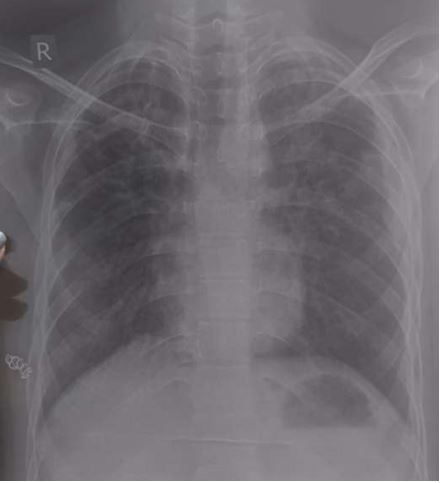
We present an interesting case report of a young non-smoker, asthmatic male with a definitive diagnosis of PLCH, clinically and radiologically. However, the classic features of Langerhans cell histiocytosis (LCH) were not seen in histopathology. We wish to highlight that advanced cases of LCH may not always have a textbook histological picture. In the appropriate clinical and radiological setting, the pathologist must consider the possibility of LCH even if classic Langerhans cells are not seen. By this paper we would like to present the features of LCH, in the advanced stage without classic presentation.
Downloads
References
2. Hidalgo A, Franquet T, Giménez A, et al. Smoking-related interstitial lung diseases: radiologic-pathologic correlation. Eur Radiol. 2006;16:2463–0.
3. Vassallo R, Limper AH, Ryu JH. Smoking-related interstitial lung disease. In: Interstitial Lung Disease, 5th ed, Schwarz MI, King TE Jr (Eds), People's Medical Publishing House, Shelton, CT, USA 2011. p.961.
4. Fernandes L, Vadala R, Mesquita AM, Vaideeswar P. Rare interstitial lung disease: Pulmonary Langerhans Cell Histiocytosis in a young non-smoking Indian female. Indian J Tuberc. 2015;62(1):46–49.
5. Deokar K, Niwas R, Chauhan N, et al. Recurrent pneumothorax, skin lesions and frequent urination. Breathe (Sheff) 2020;16(1):190318.
6. Lorillon G, Tazi A. How I manage pulmonary Langerhans cell histiocytosis. Eur Respir Rev. 2017;26:170070.
7. Suri HS, Yi ES, Nowakowski GS, Vassallo R. Pulmonary langerhans cell histiocytosis. Orphanet J Rare Dis. 2012;7:16.
8. Baqir M, Vassallo R, Maldonado F, et al. Utility of bronchoscopy in pulmonary Langerhans cell histiocytosis. J Bronchology Interv Pulmonol. 2013;20:309–2.
9. Canuet M, Kessler R, Jeung MY, Métivier AC, Chaouat A, Weitzenblum E. Correlation between high-resolution computed tomography findings and lung function in pulmonary Langerhans cell histiocytosis. Respiration. 2007;74(6):640–646.
10. Wang FF, Liu YS, Zhu WB, Liu YD, Chen Y. Pulmonary Langerhans cell histiocytosis in adults: A case report. World J Clin Cases. 2019 Jul 26;7(14):1892-1898.
11. Ninaber M, Dik H, Peters E. Complete pathological resolution of pulmonary Langerhans cell histiocytosis. Respirol Case Rep.2014;2:76–78.
12. Wolters PJ, Elicker BM. Subacute onset of pulmonary langerhans cell histiocytosis with resolution after smoking cessation. Am J Respir Crit Care Med. 2014;190:e64.
13. Lazor R, Etienne-Mastroianni B, Khouatra C, Tazi A, Cottin V, Cordier JF. Progressive diffuse pulmonary Langerhans cell histiocytosis improved by cladribine chemotherapy. Thorax.2009; 64:274–275.
Copyright (c) 2024 Author (s). Published by Siddharth Health Research and Social Welfare Society

This work is licensed under a Creative Commons Attribution 4.0 International License.

 OAI - Open Archives Initiative