
The spectrum of Spinal Cord Tumours in a Tertiary Care Centre withEmphasis on Rare Tumours: An Observational Study
Abstract
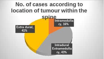
Introduction: Spinal cord tumour is an abnormal mass of tissue within/or surrounding spinal cord&/or spinal column. They are referred to according to vertebral levels and area in which they arelocated within the spine - Extradural & Intradural (Extramedullary & Intramedullary).
Aims andObjectives: To integrate histopathological spectrum of spinal cord tumours with their relevantimmunohistochemistry, their incidence and spectrum about spinal levels, location within spine, ageand sex.
Materials and Methods: Hospital-based observational study in Department of Pathology,Mahatma Gandhi Medical College, Jaipur, for two years & includes 70 cases of spinal cord tumours.
Results: In our study, 70 cases of spinal cord tumours showed 14 tumour types. Peak incidence wasseen in 61-70 years followed by 41-50 years and lowest in children ˂ 10 years. Male preponderancewas seen. The major histological type was meningiomas 17 cases (24.3%) followed byschwannomas 12 cases (17.14 %) and metastasis in 8 cases (11.4%). In relation to anatomical sitein spinal cord, tumours were most common in the thoracic spine (48.6%) followed by the cervicalspine (15.7 %).
Conclusion: In our study, the spectrum of spinal cord tumours is exhibited in 14tumour types. Peak incidence was seen in sixth decade with male preponderance and propensity forthe thoracic segment of the spine. The most common tumour type was meningiomas followed byschwannomas. IHC plays a vital role in accurate diagnosis that helps in recognizing tumourhistogenesis, clinical & radiological correlation, its pathological course, treatment & prognosis. MIB-1assesses grade & aggressiveness of tumour thus helping in evaluating its chances of recurrence.
Downloads
References
2.Gadgil NM, Chaudhari CS, Margam SR, Khan MU, Kumavat PV, Kshirsagar GR. A clinicopathological study of lesions of spinal cord and its
coverings: A tertiary care hospital experience. Annals of Pathology and Laboratory Medicine.2016 Aug10;3(3):A148-156.
3.Karsy M, Guan J, Sivakumar W, Neil JA, Schmidt MH, Mahan MA. The geneticbasis of intradural spinal tumors and its impact on clinical
treatment.Neurosurgicalfocus. 2015 Aug 1;39(2):E3.
4.Medhkour A, Chan M. Extremely rare glioblastoma multiforme of the conus medullaris with holocord and brain stem metastases, leading to cranial nerve deficit and respiratory failure: A case report and review of the literature. Surg Neurol. 2005;63:576–582. [PubMed] [Google Scholar]
5. Stein BM. Surgery of intramedullary spinal cord tumors. Clin Neurosurg. 1979;26:529–542. [PubMed] [Google Scholar]6. Ciappetta P, Salvati M, Capoccia G, Artico M, Raco A, Fortuna A. Spinal glioblastoma: report of seven cases and review of the literature. Neurosurgery. 1991;28:302–306. [PubMed] [Google Scholar]
6. B. Bouyer and N. Guedj and G. Lonjon and P. Guigui},
keywords = {Solitary fibrous tumours, Spine, Local neoplasm recurrence}, abstract = {Summary
Solitary fibrous tumours (SFTs) are rare tumours originating in the soft tissues.
7. Yadav YR, Tandon JK, Kriplani TC, Kapoor JP, Arora BM, Chandrakar S. Primary extradural spinal melanoma. Neurol India. 1995;43(3):161 -164.
8. Zileli, Mehmet & Işık, Hasan & Ogut, Fatih & Is, Merih & Cagli, Sedat & Calli, Cem. (2012). Aneurysmal bone cysts of the spine. European spine journal : official publication of the European Spine Society, the European Spinal Deformity Society, and the European Section of the Cervical Spine Research Society. 22. 10.1007/s00586-012-2510-x.
9.A. Leithner, R. Windhager, S. Lang, O. Haas, F. Kainberger, R. KotzAneurysmal bone cyst. A population based epidemiologic study and literature review.Clin. Orthop. Relat. Res., 363 (1999), Article 176–179.
10.Saavan Patel, Ryan G. Chiu, Clayton L. Rosinski, Darius Ansari, Anisse N. Chaker, Ravi S. Nunna, Mandana Behbahani, Ankit I.
Mehta,Incidence, Management, and Outcomes of Spinal Giant Cell Tumor of Bone in Adult Patients: A National Cancer Database
Analysis,World Neurosurgery,Volume 144,2020,Pages e296-e305,ISSN 1878-8750
11. Dahlin DC. Giant cell tumor of vertebrae above the sacrum: A review of 31 cases. Cancer. 1977;39:1350–6. [PubMed] [Google Scholar]
12. Kumar S, Maheshwari V, Mukherjee A, Raman DK. Primary Neuroendocrine Tumor of the Lumbar Spine: Rare Tumor Mimicking Nerve Sheath Tumor of the Spine. Asian J Neurosurg. 2019;14(3):894-896. doi:10.4103/ajns.AJNS_276_18
Copyright (c) 2022 Author (s). Published by Siddharth Health Research and Social Welfare Society

This work is licensed under a Creative Commons Attribution 4.0 International License.

 OAI - Open Archives Initiative