
A cytomorphological study of spectrum of lymphnode lesions in a tertiary care centre – retrospective study
Abstract
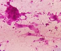
Introduction: Lymphadenopathy (LA) is a clinical condition where the lymph nodes (LNs) can be enlarged. Fine Needle Aspiration Cytology (FNAC) is a simple, inexpensive and rapid diagnostic technique practiced to find the cause for the superficial LA. With this, a study was conducted to find the cytomorphological features of the enlarged LNs and also to analyze the utility of FNAC in the suspected malignant lesions.
Materials and methods: The study was conducted in the Department of Pathology, Kamineni Institute of Medical Sciences, Narketpally, from June 2018 to Dec 2020. Random sampling was considered. Individuals of all age groups with enlarged LNs were included. Inadequate or hemorrhagic samples, non-cooperative members were not considered. The participants were proceeded for FNAC as per the standard reports. Smear preparation was done at the bedside, fixed in 95% Ethyl alcohol. The smears were stained with H & E, Giemsa techniques. If required, Ziehl Neelsen (ZN) technique was also used to find acid-fast bacilli (AFB). After staining, the smears were air-dried and examined under the microscope.
Results: A total of 161 (100%) participants were included, ages were ranged between 8 months to 85 years, and the mean age was 32.7 years. Gender wise, 89 (55.3%) were male, and 72 (44.7%) were female participants. The cervical region was identified to be the most common (108; 67%), followed by submandibular and inguinal. Among the TB LA, 8 (16%) were diagnosed to be group 1, 30 (60%) were group 2, and 12 (24%) were group 3. Out of the 22 (13.7%) malignant cases, metastasis was seen in 15 (10.55%) and lymphoma in 7 (4.34%). .
Conclusion: FNAC precludes the need for biopsy or surgery, saves time and patient resources. Hence FNAC can be considered an essential diagnostic tool in a clinical setup.
Downloads
References
Mehdi G, Singh AK, Hasan M, Ansari HA, Rehman S, Mirza S, et al. Cytological evaluation of enlarged lymph nodes in metastatic disease: A hospital-based assessment." Clinical Cancer Investigation Journal 4.2 (2015): 152.
Vatsala Sharma, Parveen Shah, Vinod Raghava, Uma Sharma. Cytomorphological Spectrum of Lymph Node Swellings: A Tertiary Care Hospital Study in Gurugram.
Vimal S, Dharwadkar A, Chandanwale SS, Vishwanathan V, Kumar H. Cytomorphological study of lymph node lesions: A study of 187 cases." Medical Journal of Dr. DY Patil University 9.1 (2016): 43.
Altınboğa, Ayşegül Aksoy, and Gökhan Yüce. Fine-Needle Aspiration Cytology in the Diagnosis of Lymph Nodes: Correlation with Histopathological Diagnosis." Southern Clinics of Istanbul Eurasia 30.1 (2019).
Ahmad T, Naeem M, Ahmad S, Samad A, Nasir A. Fine needle aspiration cytology (FNAC) and neck swellings in the surgical outpatient. J Ayub Med Coll Abbottabad. 2008 Jul-Sep; 20(3):30-2.
Iacob A, Zazgyva A, Ormenişan A, Mezei T, Sin A, Tilinca M. Effectiveness of fine-needle aspiration cytology in the diagnosis of lateral cervical nonthyroid tumors. Medicine (Baltimore). 2016 Aug; 95(31):e4448. doi: 10.1097/MD.0000000000004448.
Sohail Anwar, Amna Rehman, Asifa Karamat, Huma Batool, Ali Afzal. FNAC: FNAC: A Simple & Cost-Effective Diagnostic Tool for Benign & Malignant Pathologies Associated with Cervical Lymphadenopathy." Proceedings. Vol. 35. No. 4. 2021.
Amita S Patel, Gunvanti B Rathod, Kamlesh J Shah. Cytomorphological Study of Cervical Lymph Node Lesions. A Study of 173 cases. Ind J Pathol Res Pract. 2019; 8 (3): 255 – 62.
Hemalatha A, Shruti P, Kumar MU, Bhaskaran A. Cytomorphological patterns of tubercular lymphadenitis revisited. Ann Med Health Sci Res. 2014 May; 4(3):393-6. doi: 10.4103/2141-9248.133466.
Chaithra, S., Deoshree Solanki, and Prabhu H. Mural. Cytomorphological Study of Lymph Node Lesions: A Study of 130 Cases." Indian Journal of Pathology: Research and Practice 9.2 Part I (2020).
Ashwini HN, Mounika TS, Sneha SP, Ram SDM, Shruti Goyal S. Cytomorphological spectrum of enlarged lymph nodes. Ind J Pathol Onco. 2021; 8(2): 294 – 8. https://doi.org/10.18231/j.ijpo.2021.057
Patro P, Lad P, Hoogar MB, Reeta Dhar, Shilpi Sahu, Mithila KB, et al. Spectrum of lesions in lymph nodes-a cytological study." Int J Health Sci Res 8.11 (2018): 75-81.
Chandra TJ. One-sample two-smear versus two-sample two-smear approach for the diagnosis of pulmonary tuberculosis. J Lab Physicians. 2018 Apr-Jun; 10(2):135-139. doi: 10.4103/JLP.JLP_145_17.
Das DK, Pant JN, Chachra KL, Murthy NS, Satyanarayan L, Thankamma TC, et al. Tuberculous lymphadenitis: correlation of cellular components and necrosis in lymph-node aspirate with AFB positivity and bacillary count. Indian J Pathol Microbiol. 1990 Jan; 33(1):1-10.
Sellami M, Charfi S, Chaabouni MA, Mrabet S, Charfeddine I, Ayadi L, et al. Fine needle non-aspiration cytology for the diagnosis of cervical lymph node tuberculosis: a single center experience. Braz J Otorhinolaryngol. 2019 Sep-Oct; 85(5):617-622. doi: 10.1016/j.bjorl.2018.05.009.
Rajarikam, Nagarjuna Chary, and Lalagiri Gnana Priyanka. Fine Needle Aspiration Cytology of Lymphnode Lesions in a Rural Setup.
Copyright (c) 2022 Author (s). Published by Siddharth Health Research and Social Welfare Society

This work is licensed under a Creative Commons Attribution 4.0 International License.

 OAI - Open Archives Initiative