
A prospective report on histopathological study of ovarian tumor in a tertiary care center
Abstract
Introduction: Ovarian tumors (OTs) are the most notorious gynecological lesions in the reproductive age group. With this, a study was conducted to categorize the ovarian lesions into benign, borderline and malignant lesions and also to study their laterality, gross and microscopic patterns.
Settings: The study was conducted in the Department of Pathology, KIMS, Narketpally, Telangana from January 2017 to December 2019, 36 months. Random sampling was considered. Women aged >18yrs, histologically proven OTs, sized > 5 cm diameter, were included. Abdominal hysterectomy specimen were considered. Gross examination was done. The features such as size, the color of the specimen were noted. The stained sections were examined under a light microscope for histopathological diagnosis.
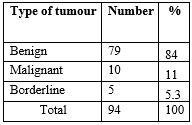
Results: Out of 94 participants, 79 were benign tumors (Bet), 5 were borderline tumour (BoT), and 10 were malignant tumor (MIT). Among the BeT, maximum were diagnosed in 21 – 40 years group, 21 – 40 years group in BoT and 41 – 60 years for MlT. In this research, 91% were unilateral, and Seromucinouscystadenoma was the common bilateral tumor. Among the different histological patterns, surface epithelial tumours constituted the majority.
Conclusion: Maximum number of tumour cases were benign, reported in the reproductive age group, whereas the malignant neoplasms in > 40 yrs. The present study emphasizes the need for proper histopathological evaluation and screening at all ages due to the relative predominance of OTs to rule out malignancies.
Downloads
References
Pachori, Geeta, et al. "Histopathological study of ovarian tumors in Ajmer region." International Journal of Medical Science and Public Health 5.7 (2016): 1400-1404.
Mankar, Deepti Vijay, and Gaurav K. Jain. "Histopathological profile of ovarian tumours: A twelve year institutional experience." Muller J Med Sci Res 6.2 (2015): 107-11.
Prat J, Mutch DG. Pathology of cancers of the female genital tract including molecular pathology. Int J Gynaecol Obstet. 2018 Oct;143 Suppl 2:93-108. doi: 10.1002/ijgo.12617.
Sudha V, Volga Harikrishnan, Sridevi. M, Padma Priya. Clinico pathological correlation of ovarian tumors in a tertiary care hospital. Ind J Of Path. Onc. 2018; 5(2): 332 – 7. https://doi.org/10.18231/2394-6792.2018.0062
Manasi, Saha, Banerjee Alpana, and Datta Abhijit. "Histological patterns of Ovarian neoplasms–A five year experience in North-East India." International Journal of Medical and Dental Sciences 7.1 (2018): 1576-1581.
Patel, Amita S., Jignasha M. Patel, and Kamlesh J. Shah. "Ovarian tumors-Incidence and histopathological spectrum in tertiary care center, Valsad." IAIM 5.2 (2018): 84-93.
Doubeni CA, Doubeni AR, Myers AE. Diagnosis and Management of Ovarian Cancer. Am Fam Physician. 2016 Jun 1;93(11):937-44.
Gurdip Kaur, Parneet Kaur, Ramesh Kumar Kundal, Jasmine Kaur. A clinicopathological study of ovarian tumors. Int J Of Cur Med Pharm Res. 2017; 3 (9): 2309 – 11. DOI: http://dx.doi.org/10.24327/23956429.ijcmpr20170225
Tamrakar, S. R., Makaju, R., Shrestha, A., & Kayastha, S. (2018). Comprehensive study of ovarian tumours in Kathmandu University Hospital. Journal of Kathmandu Medical College, 7(4), 173–179. https://doi.org/10.3126/jkmc.v7i4.23322
Mankar, Deepti Vijay, and Gaurav K. Jain. "Histopathological profile of ovarian tumours: A twelve year institutional experience." Muller J Med Sci Res 6.2 (2015): 107-11.
Kuladeepa, A. V. K., et al. "Histomorphological study of 134 primary ovarian tumors." Adv Lab Med Int 1.4 (2011): 69-82.
Manoja, Vaddadi, et al. "Clinicopathological study of ovarian tumors: a 2-year study." International Journal of Scientific Study 5.3 (2017): 297-302.
Chandanwale, Shirish S., et al. "Clinicopathologic study of malignant ovarian tumors: A study of fifty cases." Medical Journal of Dr. DY Patil University 10.5 (2017): 430.
Mondal SK, Banyopadhyay R, Nag DR, Roychowdhury S, Mondal PK, Sinha SK. Histologic pattern, bilaterality and clinical evaluation of 957 ovarian neoplasms: a 10-year study in a tertiary hospital of eastern India. J Cancer Res Ther. 2011 Oct-Dec;7(4):433-7. doi: 10.4103/0973-1482.92011.
Modepalli N, Venugopal SB. Clinicopathological Study of Surface Epithelial Tumours of the Ovary: An Institutional Study. J Clin Diagn Res. 2016 Oct;10(10):EC01-EC04. doi: 10.7860/JCDR/2016/21741.8716.
Madhumita D. K. Neeraja, Gopal A. Histomorphological spectrum of ovarian neoplasm in tertiary care centre. Int J of Recent Scientific Res. 2020; 11 (05): 38498 – 502. DOI: 10.17511/JOPM.2020.I03.06
Beulah Priscilla M, KusarajuPyla, Sindhura Manda, Satyanarayanarao P, Vijayabhaskar R. Histopathological spectrum of ovarian tumors-A three year retrospective study. IOSR J of Den and Med Sci. 2019; 3 (2): 24 – 31.
Hawaldar, Ranjana, Sadhna Sodani, and Ekta Patidar. "Histopathological spectrum of ovarian tumours-A two year retrospective study." Indian Journal of Pathology and Oncology 4.3 (2017): 450-453.
Copyright (c) 2022 Author (s). Published by Siddharth Health Research and Social Welfare Society

This work is licensed under a Creative Commons Attribution 4.0 International License.

 OAI - Open Archives Initiative