
Spectrum of Splenectomy lesions- a 12 year study at tertiary health centre
Abstract
Introduction: Splenectomy can be for a therapeutic, diagnostic, or incidental purpose. The spleencan be involved in a plethora of lesions. The purpose of the present study was to evaluatesplenectomy specimens for gross and microscopy.
Materials & Methods: An observationalretrospective-prospective study over 12 years period. All surgically resected spleens at our tertiarycare centre were evaluated for clinical, radio-laboratory investigations, gross parenchymal andmicroscopical findings.
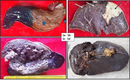
Results: Amongst 125 splenectomies, males (73.6%) and aged 20-29(34.4%) predominated. Therapeutic indications (90.4%) included laceration, splenomegaly,hypersplenism, repeated blood transfusion, splenitis and infarction. Diagnostic indications (8.0%)had cyst, abscess and lymphoma. The mean splenic weight was 480.9 grams. The highest gross-microscopy agreement related to the capsular breach, cyst, infarct and hilar vessel thrombi, whilehilar lymph node and spleniculi showed minor disagreement.
Conclusions: Meticulous grossexamination shows excellent concordance with microscopy. Diligent dissection of hilar structuresproves fruitful.
Downloads
References
1] Aster JC. Diseases of white blood cells, lymph nodes, spleen, thymus. In Kumar V, Abbas AK, Fausto M (eds) Robbins and Cotran Pathologic basis of diseases. 7th Ed. Philadelphia: Elsevier Saunders; 2005: 702- 705
2] Gupta D, Gupta A, Suri J, Singh K. Histomorphological spectrum in splenectomy specimens- A 5-year retrospective study. Indian Journal of Pathology and Oncology 2017; 4:418-421. DOI: 10.1823/2394-6792.2017.0089
3] Aliwi AJ, Al-Faddagh ZA. The Pattern of Indications for Splenectomy in Basrah. Basrah Journal of Surgery 2011; 17: 6 p.
4] Maskey P, Rupakheti S, Regmi R, Adhikary S, Agrawal CS. Splenic Epidermoid Cyst. Kathmandu University Medical Journal 2007; 5:250-2.
5] Deepankar P, Roshan R, Kumar G, Pritam A. Clinical-Etiological Profile of Patients with Splenomegaly in a Tertiary Care Centre. Journal of Medical Science and Clinical Research 2018; 6:268-274.
6] Shetty CM, Lakhkar BN, Pereira NM, Koshy SM. Role of ultrasonography and computed tomography in the evaluation of focal splenic lesions. Ind J Radiol Imag. 2005; 15:2:183-190.
7] Khalid A, UI Haque A, Naseem L. Spectrum of disease entities in splenectomy specimen. International Journal of Pathology 2006; 4: 88-93.
8] Awamleh AA, Rawabdeh S, Khasawneh H, Waqfi O, AlSoudi M. Pathological analysis of splenectomy specimens at King Hussein Medical Center: A 12-Year Study. Indian Journal of Medical Research and Pharmaceutical Sciences 2016; 3:28-32. DOI: 10.5281/zenodo.60321.
9] Vancauwenberghe T, Snoeckx A, Vanbeckevoort D, Dymarkowski S, Vanhoenackee FM. Imaging of the spleen: what the clinician needs to know. Singapore Med J. 2015; 56:133-44. DOI: 10.11622/smedj.2015040.
Copyright (c) 2022 Author (s). Published by Siddharth Health Research and Social Welfare Society

This work is licensed under a Creative Commons Attribution 4.0 International License.

 OAI - Open Archives Initiative