
A Clinicomorphological Study of Scleroderma and Morphea
Abstract
Introduction: Cutaneous involvement is a prominent feature in connective tissue disease and the skin lesions are extremely important in diagnosing and subclassifying patients with these conditions. Scleroderma (Systemic sclerosis) and Morphea (Localized Scleroderma) are two common clinical conditions with skin involvement include in connective tissue diseases of skin. Scleroderma is usually a progressive and frequently fatal disease of unknown etiology. In Morphea (Localized scleroderma) the lesions usually are limited to the skin and to the subcutaneous tissue beneath the cutaneous lesions. Although their evolutions are divergent, they share similar histologic features.
Objectives: 1.To study various histological features in cutaneous lesions of systemic scleroderma and morphea. 2. To analyze overlapping features, possible transformation of one disease to the other and co-existence in this group of diseases.
Methods: Clinically established / suspected cases of cutaneous lesions of systemic scleroderma and morphea were biopsied. After routine processing and paraffin embedding of formalin fixed tissue Haematoxyline and Eosin (H&E) sections were studied along with special stain Verhoeff vangiesson(VVG).
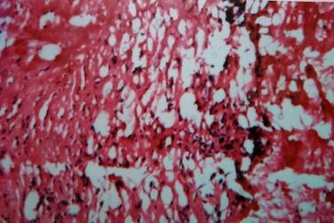
Results: Cutaneous lesions of 27 cases (9 cases of scleroderma and 18 cases of morphea) were studied and analyzed. Both the conditions were common in females.
Conclusion: Though these two conditions vary in clinical manifestations, involvement of internal organs and types of autoimmunity, the cutaneous lesions share similar histologic features. Often it is necessary to use other diagnostic tools like serological and immunological study in conjunction with histopathology with the background of clinical history for a conclusive diagnosis.
Downloads
References
2. Sappino AP, Masowye I, Saurat JH, Gabbian’s G. Smooth muscle differentiation in Scleroderma fibroblastic cells. American Journal of Pathology 1990; 137: 585-591.
3. D’Angelo WA, Fries JF, Masi AT, Shulman LE. Pathologic observations in systemic sclerosis (Scleroderma). A study of fifty-eight autopsy cases and fifty-eight matched controls. Am J Med. 1969 Mar: 46(3):428-440.
4. Tremaine R, Adam JE, Orizaga M. Morphea coexisting with lichen sclerosus et atrophicus. Int J Dermatol. 1990 Sep;29(7):486-9. [PubMed]
5. Fleischmajer R, Damiano V, Nedwich A. Alteration of subcutaneous tissue in systemic scleroderma. Arch Dermatol. 1972 Jan;105(1):59-66. [PubMed]
6. Leinwand I,Duryee AW, Richter MN. Scleroderma; based on a study of over 150 cases. Ann Intern Med. 1954 Nov;41(5):1003-41.
7. Shono S, Imura M, Ota M, Osaku A, Shinomiya S, Toda K. Lichen sclerosus et atrophicus, morphea, and coexistence of both diseases. Histological studies using lectins. Arch Dermatol. 1991 Sep;127(9):1352-6.
8. Lever WF. Histopathology of the skin. J.B. Lippincott, Philadelphia, 1949.
9. Jaworsky C. Connective tissue diseases in Histopathology of the skin. Philadelphia; Lippincott Raven, 8th edition, 1997. [PubMed]
10. O’Leary PA, Montgomery H, Ragsdale WE. Dermatohistopathology of various types of scleroderma. AMA Arch Derm. 1957 Jan;75(1):78-87. [PubMed]

 OAI - Open Archives Initiative