
The spectrum of palpable breast lesions- A cytopathological study of 1193 cases
Abstract
Introduction- The vast majority of the lesions that occur in the breast are benign. Much concern is given to malignant lesions of the breast because female breast cancer has now surpassed lung cancer as the leading cause of global cancer incidence in 2020. Fine needle aspiration cytology (FNAC) has good sensitivity, specificity and accuracy in the diagnosis of both neoplastic and non-neoplastic breast lump thereby assisting in early diagnosis and further management. The current study was done to study the incidence and the different cytomorphological patterns of palpable breast lumps by FNAC and consequently compare the results with studies in the literature.
Materials and methods- This is a retrospective study conducted from January 2018 to December 2020 in a tertiary care hospital. The three-year data is obtained from the records maintained in the FNA clinic. The patients were counselled before the procedure and informed consent was taken.
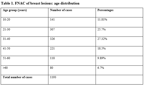
Results- A total of 1193 breast lump cases were analysed in this 3-year study, there were 19 male patients all of them presented with gynaecomastia and 1177 female patients. The patient’s age group ranged from 12 to 86 years. The commonest age group with the lesions 31-40 years comprising 326 cases (27.32%) followed 21-30 years age group in the second place with 307 cases (25.7%).
Conclusion- In this study the most common benign neoplastic and malignant neoplastic breast lumps are fibroadenoma and infiltrating ductal carcinoma respectively. Fibrocystic disease of the breast is the most common non-neoplastic breast lump.
Downloads
References
Bedrossian CW. Bridging the gap between cytopathology and surgical pathology. Diagn Cytopathol. 1995 Feb;12(1):1-2. doi: 10.1002/dc.2840120102.
Godwins, Echejoh, Dzuachi David, and Jenrola Akeem. "Histopathologic analysis of benign breast diseases in Makurdi, North Central Nigeria." International Journal of Medicine and Medical Sciences 3.5 (2011): 125-128.
Guray M, Sahin AA. Benign breast diseases: classification, diagnosis, and management. Oncologist. 2006 May;11(5):435-49. doi: 10.1634/theoncologist.11-5-435.
Sung H, Ferlay J, Siegel RL, Laversanne M, Soerjomataram I, Jemal A, et al. Global Cancer Statistics 2020: GLOBOCAN Estimates of Incidence and Mortality Worldwide for 36 Cancers in 185 Countries. CA Cancer J Clin. 2021 May;71(3):209-249. doi: 10.3322/caac.21660.
Gupta S. Breast cancer: Indian experience, data, and evidence. South Asian J Cancer. 2016 Jul-Sep;5(3):85-6. doi: 10.4103/2278-330X.187552.
Malvia S, Bagadi SA, Dubey US, Saxena S. Epidemiology of breast cancer in Indian women. Asia Pac J Clin Oncol. 2017 Aug;13(4):289-295. doi: 10.1111/ajco.12661.
Anders CK, Hsu DS, Broadwater G, Acharya CR, Foekens JA, Zhang Y, et al. Young age at diagnosis correlates with worse prognosis and defines a subset of breast cancers with shared patterns of gene expression. J Clin Oncol. 2008 Jul 10;26(20):3324-30. doi: 10.1200/JCO.2007.14.2471. Erratum in: J Clin Oncol. 2011 Sep 20;29(27):3721.
Koss, L. "Diagnostic cytology 4th edition." (1992): p6-11.
Orell, Svante R., Gregory F. Sterrett, and Daniel Whitaker. Fine needle aspiration cytology. Elsevier Churchill Livingstone, 2005.
Chandanwale SS, Gupta K, Dharwadkar AA, Pal S, Buch AC, Mishra N. Pattern of palpable breast lesions on fine needle aspiration: A retrospective analysis of 902 cases. J Midlife Health. 2014 Oct;5(4):186-91. doi: 10.4103/0976-7800.145164.
Rachana B, Shweta D, Mayur A, Grace DC. Cytomorphological spectrum of breast lesions diagnosed by fine needle aspiration cytology. International Journal of Medical and Health Research. 2018; 4(8):168-71.
Likhar, K. S., Fatima, A., Hazari, R. A., Gupta, S. G., & Shukla, U. (2013). Diagnostic role of FNAC in breast lesions. IJRRMS, 3(1), 12-4.
Kamra, H. T., Rana, P., Kaur, S., Verma, S., Munde, S., et al. Spectrum of breast lesions diagnosed on fine needle aspiration cytology in rural population of Khanpur Kalan, Sonepat (Haryana). Ann Int Med Den Res, 3(3), 06-09.
Akhator, Afeyodion. "Benign breast masses in Nigeria." Nieg Jr of Surg Sciences 17 (2007): 105-8.
Irabor, D. O., and C. A. Okolo. "An audit of 149 consecutive breast biopsies in Ibadan, Nigeria." Pakistan Journal of Medical Sciences 24.2 (2008): 257.
Khan, A., Jamali, R., Jan, M., & Tasneem, M. (2014). Correlation of fine needle aspiration cytology and histopathology diagnosis in the evaluation of breast lumps. International Journal of Medical Students, 2(2), 40-43.
Rahman MZ, Sikder AM, Nabi SR. Diagnosis of breast lump by fine needle aspiration cytology and mammography. Mymensingh Med J. 2011 Oct;20(4):658-64.
Chhieng DC, Fernandez G, Cangiarella JF, Cohen JM, Waisman J, Harris MN, et al. Invasive carcinoma in clinically suspicious breast masses diagnosed as adenocarcinoma by fine-needle aspiration. Cancer. 2000 Apr 25;90(2):96-101.
Ballo MS, Sneige N. Can core needle biopsy replace fine-needle aspiration cytology in the diagnosis of palpable breast carcinoma. A comparative study of 124 women. Cancer. 1996 Aug 15;78(4):773-7. doi: 10.1002/(SICI)1097-0142(19960815)78:4<773::AID-CNCR13>3.0.CO;2-S.
Copyright (c) 2021 Author (s). Published by Siddharth Health Research and Social Welfare Society

This work is licensed under a Creative Commons Attribution 4.0 International License.

 OAI - Open Archives Initiative