
Cell pattern abnormalities in cervical pap smear in correlation with age and demography at a Tertiary care centre.
Abstract
Introduction: Carcinoma Cervix is common all around the globe and ranked third amidst all malignancies among women. The cervical mucosa undergoes morphologic variation with age and practising cytopathologists is aware of these difference to make an accurate diagnosis. This study aimed to detect abnormal cervical epithelial cell patterns in a rural population and compare lesions or abnormal cell patterns among different age groups.
Materials and Methods: This is a cross-sectional, descriptive study conducted in a tertiary care centre at the Department of Pathology over 6 months. 408 women were included in the study. Data were entered in Microsoft Excel and analyzed in SPSS software.
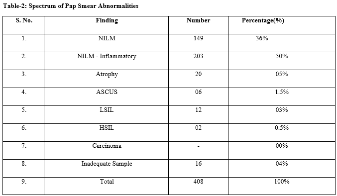
Results: Out of 408 women included in the study, the most common age group of the presentation was 31 to 40 years (36%), followed by 20 to 30 years (24%). NILM-Inflammatory was the most common finding (50%), followed by NILM (36%). The most common symptoms of presentation were Menstrual abnormalities (21%), White discharge and pruritus (18%). Findings in younger women were most commonly NILM-Inflammatory & NILM whereas in the post-menopausal age group, ASCUS, LSIL & HSIL.
Conclusion: All women above 30 years of age should undergo routine cervical cancer screening, and should continue screening even in the perimenopausal and postmenopausal age. Most women who visited the gynaecology OPD were not aware of cervical cancer screening. Hence the general population has to be educated about the benefits of pap smear test through medical camps and awareness programs.
Downloads
References
Tamboli G.D. et al. Accuracy of cytological findings in abnormal cervical smear by cyto-histological comparison. J Med Educat Res. 2013;3(2):19-24.
Banik U, Bhattacharjee P, Ahamad SU, Rahman Z. Pattern of epithelial cell abnormality in Pap smear: A clinicopathological and demographic correlation. Cyto Journal 2011;8:8. doi: 10.4103/1742-6413.80527.
Adhikary AK, Banik U, Numaga J, Suzuki E, Inada T, Okabe N. Heterogeneity of the fibre sequence in subgenus C adenoviruses. J Clin Pathol 2004;57(6):612-7. doi: 10.1136/jcp.2003.014944.
Toews HA. The abnormal pap smear: A rationale for follow up. Can Fam Physician. 1983;29:759-62.
Elmslie TJ. The pap smear and cervical cancer screening. Can Fam Physician 1987;33:131-7.
Chhieng DC, Roberson J, Gidley J, Eltoum I. Bethesda 2001. Impact on the reporting of gynecologic cytology. Acta Cytol 2004;48:355-62
Stewart BW, Kleihues P. World Cancer Report. Lyon: IARC Press; 2003.
Suba EJ, Raab SS. Viet/American Cervical Cancer Prevention Project. Papanicolaou screening in developing countries: An idea whose time has come. Am J Clin Pathol 2004;121:315-20.
Suba EJ, Murphy SK, Donnelly AD, Furia LM, Huynh ML, Raab SS. Systems analysis of real-world obstacles to successful cervical cancer prevention in developing countries. Am J Public Health 2006;96:480-7.
Pun RG, Shrestha J, Awale PJ, Chitrakar N, Jha R, Khadka SS. Cytological pattern of cervical pap smears. J Pathol Nep 2018;8:1280-4.
Das D, Kar A, Rath S, Baliarsingh SK, Prusty D, Dash AK. Cytological pattern of Papanicolaou smears and detection of cervical cancers: An experience from a tertiary care centre of eastern zone of India. Oncol J India 2018;2:25-8
Atla BL, Uma P, Shamili M, Kumar SS. Cytological patterns of cervical pap smears with histopathological correlation. Int J Res Med Sci 2015;3(8):1911-6.
Sachan PL, Singh M, Patel ML, Sachan R. A Study on Cervical Cancer Screening Using Pap Smear Test and Clinical Correlation. Asia Pac J Oncol Nurs 2018;5:337-41.
Aytekin Tokmak1, Ali Irfan Guzel1, Emre Ozgu, Murat Oz, Serap Akbay, Salim Erkaya, Tayfun Gungor. Clinical Significance of Atypical Squamous Cells of Undetermined Significance in Detecting Preinvasive Cervical Lesions in Post- Menopausal Turkish Women.Asian Pac J Cancer Prev, 15 (16), 6639-6641.
Akshatha C, Arul P, Shetty S. Prevalence and comparison of cervical cytology abnormalities in postmenopausal and elderly women: A experience from tertiary care hospital. J MedSoc 2017;31:23-7.
Misra JS, Srivastava AN, Zaidi ZH. Cervical cytopathological changes associated with onset of menopause. J Mid-life Health 2018;9:180-4.
Kaustubh Mulay, Meenakshi Swain, Sushma Patra SG. A comparative study of cervical smears in an urban Hospital in India and a population-based screening program in MauritiusNo Title. Indian J Pathol Microbiol. 2009;52:34–7.
Bhavika K. VaghelaVKV and PMS. Analysis of abnormal cervical cytology in Papanicolaou smears at tertiary care center – A retrospective study. Int J Biomed Adv Res. 2014;5:47–9.
Copyright (c) 2021 Author (s). Published by Siddharth Health Research and Social Welfare Society

This work is licensed under a Creative Commons Attribution 4.0 International License.

 OAI - Open Archives Initiative