
Histopathological spectrum of Cervical Lesion”– two and half Year prospective Study in Tertiary Care Center of Chhattisgarh, India
Abstract
Introduction: Cervical lesions are the leading cause of morbidity in Indian women and cervical cancer is the second most common cancer in women worldwide next to breast cancer.
Objectives: To study the age distribution, the relative frequency of various cervical lesions and histopathological features of cervical lesions.
Materials and Methods: This is a two & half years retrospective study of all cervical biopsies and hysterectomy specimens received from September 2017 to March 2020 in the department of pathology.
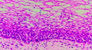
Result: In a total of 485 cases studied 359 (74.1%) cases were non-neoplastic, 107(22%) were preinvasive and 19 (3.9%) cases were malignant. Cervicitis was the most common non-neoplastic lesion and squamous cell carcinoma was the most common cancer.
Conclusion: Our study highlights a vast spectrum of cervical lesions and therefore early detection and management of certain lesions can help in reducing morbidity.
Downloads
References
Bray F, Ferlay J, Soerjomataram I, Siegel RL, Torre LA, Jemal A. Global Cancer Statistics 2018: GLOBOCAN estimates of incidence and mortality worldwide for 36 cancers in 185 countries. CA Cancer J Clin, in press. The online GLOBOCAN 2018 database is accessible at http://gco.iarc.fr/, as part of IARC’s Global Cancer Observatory.
Srivani Saravanan, Jonathan Arnold, Arul P. “Histomorphological Spectrum of Lesions of the Cervix, A Retrospective Study in a Tertiary Care Hospital”. Journal of Evolution of Medical and Dental Sciences 2015; July 4(59) : 10326-10329.
Bosch FX, Lorincz A, Muñoz N, Meijer CJ, Shah KV. The causal relation between human papillomavirus and cervical cancer. J Clin Pathol. 2002 Apr;55(4):244- 65.
Fritz A, Percy C, Jack A, Shanmugaratnam K,Sobin LH, Parkin DM, Whelan S, International classification of Diseases for oncology (ICD-0), 3rd edition, World health organization: Geneva, 2000.
Krishnappa C, Kanabur DR, Dinesh CU. Clinico-morphological Spectrum of Neoplasms of Uterine Cervix in a Tertiary Care Center in North Karnataka, South India. Int J Sci Stud 2016;4(2):6-12.
Pradhan B, Pradhan SB, Mital VP. Correlation of PAP smear findings with clinical findings and cervical biopsy. Kathmandu Univ Med J (KUMJ) 2007;5:461-7.
Shruthi PS, Kalyani R, Kai LJ, Narayanaswamy M. Clinicopathological correlation of cervical carcinoma: A tertiary hospital based study. Asian Pac J Cancer Prev 2014;15:1671-74.
Fotra R, Gupta S, Gupta S. Sociodemographic risk factors for cervical cancer in Jammu region of J and K state of India first ever report from Jammu. Indian J Sci Res 2014;9:105-10.
Sinha P, Rekha PR, Subramaniam PM, Konapur PG, Thamilselvi R, Jyothi BL. A Clinicomorphological study of carcinoma cervix. Nat J Basic Med Sci 2011;2:2-7.
Jashamy KA, Al-Naggar RA, San P, Mashani M. Histopathological findings for cervical lesions in Malaysian women. Asian Pac J Cancer Prev 2009;10:1159-62.
Dr. BVVD Kiranmayi. “Morphological Spectrum of Cervical lesions with an emphasis on Neoplastic lesions - a 2year retrospective study.” IOSR Journal of Dental and Medical Sciences (IOSR-JDMS) , vol. 16, no. 11, 2017, pp. 54–57.
Bagde, Gupta R, Ganguly S, Bhardwaj A, Jogis. “Spectrum of Cervical Lesions in CIMS, Bilaspur: A 5 years Retrospective Study of 215 Cases in a Tertiary Hospital of Central India” Journal of Evidence based Medicine and Healthcare. 2015 Oct19; 2(42): 7505-7510
Nwachokor FN, Forae GC. Morphological spectrum of non-neoplastic lesions of the uterine cervix in Warri, south-south, Nigeria. Niger J ClinPract. 2013 Oct-Dec; 16(4):429-32.doi: 10.4103/1119-3077.116883.
Richards MJ, Angus D. Possible sexual transmission of genitourinary tuberculosis. Int J Tuberc Lung Dis 1998; 2:439. M
oussa B, valentine K,Adama O,Aziz D A, Idrissa Z, Goumburi LO (2016). Tuberculosis of the Uterine Cervix: About a Case and Literature Review. Open Journal of Obstetrics and Gynecology .6,734-739.
Hatwal D, Batra N, Kumar A, Chaudhari S, Bhatt S. Spectrum of Nonneoplastic Lesions of Uterine Cervix in Uttarakhand. National Journal of Laboratory Medicine. (Feb.)2016;1-5. DOI: NJLM/2016/18005:2098.
Bansal A, Kumar A, Reddy GT. Benign lesions of cervix uteri: without human papilloma virus. International Journal of Research and Review. 2019; 6(11):254-259.
Tamboli GD, Khatod LV. Accuracy of cytological findings in abnormal cervical smear by cyto-histological comparision. J Medical Education Research. 2013;3(2):19-24.
Saha R, Thapa M. Correlation of Cervical cytology with Cervical Histology. Kathmandu University Med J Kumj. 2015;3:222-4.
Kumari k,Umrani M.K.,bharathi M. Histopathological spectrum of cervical biopsies- a 5 year retrospective study.Trop J path micro 2017;3(1):46-51.doi:10.17511/jopm.2017.i1;08.
Alfsen GC, Kristensen GB, Skovlund E, Pettersen EO, Abeler VM. Histologic subtype has minor importance for overall survival in patients with adenocarcinoma of the uterine cervix: A population-based study of prognostic factors in 505 patients with nonsquamous cell carcinomas of the cervix. Cancer 2001;92:2471-83.
Jain A, Dhar R, Patro P et.al. Histopathological study of cervical lesions. Int J Health Sci Res. 2018; 8(11):82-87
Krishna T et al., Sch. J. App. Med. Sci., Apr 2017; 5(4C):1443-1449
Shingleton HM, Bell MC, Fremgen A, Chmiel JS, Russell AH, Jones WB, et al. Is there really a difference in survival of women with squamous cell carcinoma, adenocarcinoma, and adenosquamous cell carcinoma of the cervix? Cancer 1995;76:1948-55.
Galic V, Herzog TJ, Lewin SN, Neugut AI, Burke WM, Lu YS, et al. Prognostic significance of adenocarcinoma histology in women with cervical cancer. Gynecol Oncol 2012;125:287-91.
Jeong BK, Choi DH, Huh SJ, Park W, Bae DS, Kim BG. The role of squamous cell carcinoma antigen as a prognostic and predictive factor in carcinoma of uterine cervix. Radiat Oncol J 2011;29:191-8.
Galic V, Herzog TJ, Lewin SN, Neugut AI, Burke WM, Lu YS, et al. Prognostic significance of adenocarcinoma histology in women with cervical cancer. Gynecol Oncol 2012;125:287-91.
Copyright (c) 2021 Author (s). Published by Siddharth Health Research and Social Welfare Society

This work is licensed under a Creative Commons Attribution 4.0 International License.

 OAI - Open Archives Initiative