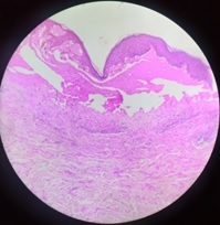
Histopathological study of non-neoplastic skin lesions in a tertiary care center
Abstract
Introduction: Skin is the largest organ of our body. Non-neoplastic skin lesions are more common than neoplastic lesions. The histopathological study was done to know the prevalence of various non-neoplastic skin lesions of patients who attended the outpatient department of dermatology over a period of three years from Jan 2016-Dec2018. The present study was based on the histopathological presentation of various non-neoplastic skin lesions, their prevalence, and classifying the lesions into various categories.
Materials and Methods: In this study total of 209 cases of skin lesions were taken over a period of three years. The diagnosis of these skin lesions was confirmed by histopathological examination with routine hematoxylin and eosin stain.
Results: A total of 209 cases of non-neoplastic lesions were taken for the study. Out of these lesions, 63 cases (30.14 %) were non-infectious - vesiculobullous, 54 ( 25.84 %) were reported under the category of infectious etiology, 41 cases (19.62 %) of non-infectious erythematous papulosquamous diseases, 13 cases (6.22 %) of inflammatory disorders, 10 (4.78 %) cases showed connective tissue disorders. 8(3.83%) cases were reported as vasculitis and 2 cases (0.96) of fungal origin. 18 cases come under the miscellaneous category that was correlated clinically and were treated.
Conclusion: In the present study of non-neoplastic skin lesions, non-infectious vesiculobullous diseases were more common. Pemphigus Vulgaris was the most common lesion. The non-neoplastic skin lesions were most commonly seen in males than females in our population of the study.
Downloads
References
Kumar V, Goswami HM. Spectrum of non-neoplastic skin lesions: a histopathological study based on punch biopsy. Int J Cur Res Rev. 2018; 10(6):43-48. doi:10.7324/IJCRR.2018.1069.
Narang S, Jain R. An evaluation of histopathological findings of skin biopsies in various skin disorders. Ann Pathol Lab Med. 2015;02(01):42-46.
Mehar R, Jain R, Kulkarni CV, Narang S, Mittal M, Patidar H. Histopathological study of dermatological lesions – A retrospective approach. Int J Med Sci Public Health. 2014;3(9):1082-1085. doi: 10.5455/ijmsph.2014.190620142.
Goyal N, Jain P, Malik R, Koshti A. Spectrum of Non Neoplastic Skin Diseases: A Histopathology Based Clinicopathological Correlation Study. Sch J App Med Sci. 2015;3(1F):444-449.
Achalkar GV. Clinico-pathological evaluation of non-neoplastic and neoplastic skin lesions: A study of 100 cases. Indian J Pathol Oncol. 2019;6(1):118-122. doi: 10.18231/2394-6792.2019.0021.
Yahya H. Change in pattern of skin disease in Kaduna, North-Central Nigeria. Int J Dermatol. 2007;46(9):936-43. doi: 10.1111/j.1365-4632.2007.03218.
Ogunbiyi AO, Owoaje E, Ndahi A. Prevalence of skin disorders in school children in Ibadan, Nigeria. Pediatr Dermatol. 2005;22(1):6-10. doi: 10.1111/j.1525-1470.2005.22101.x.
R Singh, K Bharathi, R Bhat, C Udayashankar. The Histopathological Profile of Non-Neoplastic Dermatological Disorders with Special Reference to Granulomatous Lesions - Study At A Tertiary Care Centre In Pondicherry. Int J Pathol. 2012;13(3):1-6.
Agarwal D, Singh K, Saluja KS, Kundu PR, Kamra H, Agarwal A. Histopathological Review of Dermatological Disorders with a Keynote to Granulomatous Lesions: A Retrospective Study. Int J Sci Study. 2015;3(9) 66-69. doi: 10.17354/ijss/2015/557.
Gautam K, Pai RR, Bhat S. Granulomatous lesions of the skin. J Pathol Nepal. 2011;1(2):81-86. doi: 10.3126/jpn.v1i2.5397.
Veldurthy VS, Shanmugam C, Sudhir N, Sirisha O, Motupalli CP, Rao N, Reddy SR, Rao N. Pathological study of non-neoplastic skin lesions by punch biopsy. Int J Res Med Sci. 2015;3(8):1985-1988. doi: 10.18203/2320-6012.ijrms20150313.
Chavhan SD, Mahajan SV, Vankudre AJ. A Descriptive Study on Patients of Papulosquamous Lesion at Tertiary Care Institute. MVP J Med Sci. 2014;1(1):30-35.

Copyright (c) 2020 Author (s). Published by Siddharth Health Research and Social Welfare Society

This work is licensed under a Creative Commons Attribution 4.0 International License.


 OAI - Open Archives Initiative


