
A demographic study of a series of eosinophilic appendicitis and its correlation with the clinicopathological profile in a tertiary care hospital-a 3-year study
Abstract
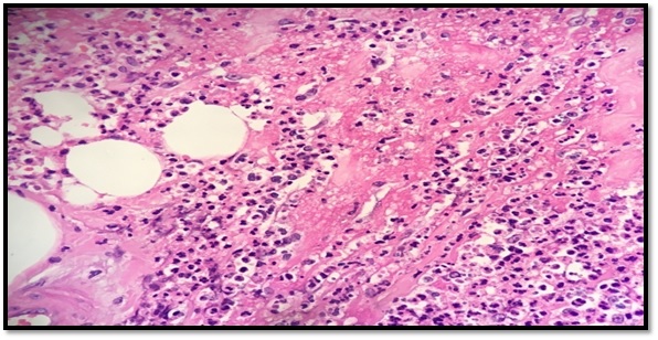
Introduction: Acute eosinophilic appendicitis (AEA) is a rare variant of inflammation of the appendix. The histologic hallmark of this entity is the infiltration of eosinophils in the musularis layer of the appendix with edema separating the muscle fibers with very few or no neutrophilic infiltration. A retrospective study was done to determine their demographic profile, morphological diagnosis, and clinical correlation.
Materials and Methods: All cases of Acute appendicitis reported at the histopathology department of Sri Muthukumaran Medical College and Research Institute, a teaching hospital from the year of Jan 2017 to Dec 2019 were included in this study. Histopathological examination is the gold standard for the diagnosis of acute appendicitis. Hence, Formalin-fixed paraffin-embedded tissue which was stained with Hematoxylin and Eosin were retrieved and used for routine morphology and diagnosis.
Results: Out of 419 cases of appendicectomies studied, 27 (6.49%) cases were found to be acute eosinophilic appendicitis, 265 (63.7%) cases were acute appendicitis, 30 (7.2%) cases were reported as acute suppurative appendicitis with periappendicitis most of which showed infiltration of eosinophils along with other inflammatory cells and 97 (23.3%) cases were reported as appendix with reactive lymphoid hyperplasia. Most of the acute appendicitis showed gross edema and congested blood vessels on the serosal surface. Enterobius vermicularis worm infestation was noted in 7 (25%) cases reported as acute eosinophilic appendicitis and in 20 (7.5%) cases of acute appendicitis.
Conclusion: It was inferred that the acute eosinophilic appendicitis is a condition which could be an allergic response and an early event in the evolution of acute phlegmonous appendicitis.
Downloads
References
Liu C, Crawford JM. The Gastrointestinal tract. In: Kumar V, Abbas AK, Fausto N, eds. Robbins and Cotran Pathologic Basis of Disease. 7th edition. Philadelphia PA: Elsevier inc.; 2004. p. 797-875.
Barcia JJ, Reissenweber N. Neutrophil count in the normal appendix and early appendicitis: Diagnostic index of real acute inflammation. Ann Diagn Pathol. 2002;6(6):352-356. doi: https://doi.org/10.1053/adpa.2002.36659.
. Pieper R, Kager L, Näsman P. Clinical significance of mucosal inflammation of the vermiform appendix. Ann Surg 1983;197(3):368-374. doi: https://dx.doi.org/10.1097%2F00000658-198303000-00019.
Norman J, Leslie H. Appendix. In: Weidner N, Cote R, Suster S, Weiss LM, Modern surgical pathology. 2nd ed. Saunders Elsevier. 2009 vol.1. pp. 837-852
Carr NJ. The pathology of acute appendicitis. Ann Diag Pathol 2000;4(1):46-58. doi: https://doi.org/10.1016/s1092-9134(00)90011-x.
Gaurav Jain, Sujata R Kanetkar. Eosinophilic appendicitis: Case report of five cases & review of literature. J Med Res Pract. 2013;2(8):208-211.
Aravindan KP, Vijayaraghavan D, Manipadam MT. Acute eosinophilic appendicitis and the significance of eosinophil-Edema lesion. Indian J Pathol Microbiol. 2010;53(2):258-261. doi: https://doi.org/10.4103/0377-4929.64343.
Stemmerman GN. Eosinophilic granuloma of the appendix: a study of its relation to Strongyloides infestation. Am J Clin Pathol. 1961;36:524-531. doi: https://doi.org/10.1093/ajcp/36.6.524.
Tally NJ, Shroter RG, Philips SF, Zinsmeister AR. Eosinophilic gastroenteritis: a clinicopathological study of patients with disease of the mucosa, muscle layer and the subserosal tissue. Gut.1990:31(1):54-58. doi: https://doi.org/10.1136/gut.31.1.54.
Shivraj N Kanthikar, Dhiraj B Nikumbh, Sneha S Desale: Primary Eosinophilic Obliterative Appendicitis: Online J Health All Sci. 2014;13(1):6.
Stephenson J, Snoddy WT. Appendiceal lesions: Observation in 4000 appendectomies. Arch Surg. 1961;83(5): 661-666. doi: https://doi.org/10.1001/archsurg.1961.01300170017005.
Rothenberg ME. Eosinophilic gastrointestinal disorders EGID). J Allergy Clin Immunol. 2004;113(1):11-28. doi: https://doi.org/10.1016/j.jaci.2003.10.047.
Alvarado A: A practical score for the early diagnosis of acute appendicitis. Ann Emerg Med. 1986;15(5):557-564. doi: https://doi.org/10.1016/s0196-0644(86)80993-3.
Shrestha R, Ranabhat SR, Tiwari M. Histopathologic analysis of appendectomy specimens. J Pathol Nepal. 2012;2(3):215-219. doi: https://doi.org/10.3126/jpn.v2i3.6025.
Bickell NA, Aufses Jr AH, Rojas M, Bodian C. How time affects the risk of rupture in appendicitis. J Am Coll Surg. 2006;202(3):401-406. doi: https://doi.org/10.1016/j.jamcollsurg.2005.11.016.
Bhangu A, Søreide K, Di Saverio S, Assarsson JH, Drake FT. Acute appendicitis: modern understanding of pathogenesis, diagnosis, and management. The Lancet. 2015;386(10000):1278-1287. doi: https://doi.org/10.1016/S0140-6736(15)00275-5.
Sinha RT, Dey A. A retrospective study of histopathological features of appendectomy specimens–what all can expect. J Med Sci Health. 2016;2(2):6-12.
Polat DA, Münevver M, Selçuk U, MahirM O, VasfiM O, Selda S, Faruk C. Unusual findings in appendectomy specimens: Evaluation of 2458 cases and review of the literature. Indian Journal of Surgery. 2004;66(4):221-226.
Liu, C., Crawford, J.M. 2004. The Gastrinitestinal tract. In: Kumar V, Abbas AK,Fausto N, eds. Robbins and Cotran Pathologic Basis of Disease. 7th edition. Philadelphia PA: Elsevier inc.; p. 797-875.
Blackshaw AJ, Levison DA. Eosinophilic infiltrates of the gastrointestinal tract. J Clin Pathol. 1986;39(1):1-7. doi: https://dx.doi.org/10.1136%2Fjcp.39.1.1.
Copyright (c) 2020 Author (s). Published by Siddharth Health Research and Social Welfare Society

This work is licensed under a Creative Commons Attribution 4.0 International License.

 OAI - Open Archives Initiative