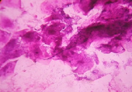
Cytologic Manifestations of Vaginitis
Abstract
Introduction: As the lower genital tract is directly exposed to the external environment, it is subjected to inflammation as well as infection, which may remain localized or may progress to other areas such as the endometrium, fallopian tubes, peritoneal cavity and, less likely, the ovaries. Vaginitis is a common health problem. The main aim of the present study is to recognize the cytological manifestations in vaginitis and to identify the possible causative agent.
Materials and Methods: Cytologic evaluations of vaginal smear were made in 167 patients. Clinical details were obtained by examining the patients and relevant investigations were noted. With the consent of the patient posterior vaginal fornix was swabbed with cotton tipped applicator. PH was determined. Whiff’s amine test was done for presence of fishy amine odor. Wet mount preparation was immediately made and examined under microscope. Three smears were made, one each for Gram’s, Giemsa and papanicolaou.
Result: Out of 167 cases, most common cytological diagnoses offered were Bacterial vaginosis (50.8%), Koilocytotic atypia (13.7%), Trichomonas vaginalis infestations (12.5%), and vaginal candidiasis (4.8%). Majority were in age group 21-30 years (49.59%).
Conclusion: There were diverse cytologic manifestations in vaginitis and was useful in identifying the possible etiological factors. Hence the results obtained are useful in screening large population.
Downloads
References
2. Edwards L. Vaginitis. In : Black M, Rudolph CMA, Edward L, Lynch PJ. Obstetric and gynaecological dermatology. 3rd edn. Mosby: Elsevier; 2008:301-16.
3. Ainbinder SW, Ramin SM, DeCherney AH. Sexually transmitted diseases and pelvic infections. In : DeCherney AH, Nathan L, Goodwin TM, Laufer Neri. Current Diagnosis and treatment Obstetric and Gynecology. 10th edn. McGraw Hill: New York, 2003;662-95.
4. Boon ME, Gray W. Normal Vulva, vagina and cervix : hormonal and inflammatory conditions. In : Gray W, McKee GT, editors. Diagnostic cytopathology. 2nd ed. Philadelphia : Churchill Livingstone : 2003:651-705.
5. Amsel R, Totten PA, Spiegel CA, Chen KC, Eschenbach D, Holmes KK. Nonspecific vaginitis. Diagnostic criteria and microbial and epidemiologic associations. Am J Med. 1983 Jan;74(1):14-22.
6. Koss LG, Melamed MR editors. Benign disorders of uterine cervix and vagina. In: Koss’ diagnostic cytology and its histopathologic basees. 5th ed. Vol.1, Philadelphia: Lippincott Williams and Wilkins; 2006:241-81.
7. Henry MR. The Bethesda system 2001: an update of new terminology for gynaecologic cytology. Clinics in laboratory medicine 2003 Sep;23(3):585-603.
8. Sobel IO. Vaginitis. N Engl J Med 1997;337(26):1896-1903. [PubMed]
9. David C, Foster. Vulvitis and Vaginitis. Current Opinion in Obstet & gynecol 1993,5 :726-32. [PubMed]
10. Kiviat NB, Paavonen JA, Brockway J, Critchlow CW, Brunham RC, Stevens CE, Stamm WE et al. Cytologic manifestations of cervical and vaginal infections I. Epithelial and inflammatory cellular changes. JAMA 1985;253(7):989-96.
11. Schnadig VS, Davie KD, Shafer SK, Yandell RB, Islam MZ, Hannigan EV. The cytologist and bacterioses of the Vaginal – ectocervical area clues, commas and confusion. Acta Cytol 1989;33(3):287-97. [PubMed]
12 . Herrero R, Munoz N, et al. HPV International Prevalence Surveys in general population. (Abstract) 18 International HPV Conference. Barcelona. 2000:126.
13. Sonnex C, Lefor W. Microscopic features of vaginal candidiasis and their relation to symptomalotogy Sex Transm Inf 1999;75:417-9. [PubMed]
14. Schwebke JR, Burgess D. Trichomonas. Clin Microbial Rev. 2004:17(4):794-802. [PubMed]
15. Demirezen S, Safi Z, Beksac S. The interaction of trichomonas vaginatis with epithelial cells, polymorphonuclear leucocytes and erythrocytes on vaginal smears; light microscopin observation. Cytopathol 2000;11:326-333.
16. Bhagawan BS, Gupta PK. Genital actinomycosis and intrauterine contraceptive devices – cytopathologic diagnosis and clinical significance. Hum Pathol 1978;9(5):567-78.
17. Panikabutra K. Clinical aspects of uncomplicated gonorrheae in the female. Br J Vener Dis 1973;49(2): 213-15. [PubMed]
18. Richens j.The diagnosis and treatment of donovanosis (granuloma inguinale). Genitourin Med 1991;67:441-452. [PubMed]


 OAI - Open Archives Initiative


