
Diagnosis of primary site of tumour origin using immunohistochemistry on cell block in cases of malignant ascitic fluid
Abstract
Introduction: Presence of malignant cells in peritoneal fluid has serious implications as most of the times, it represents a metastasis from a distant organ, along with poor prognosis.
Aim: 1) To ascertain primary site of origin from malignant ascitic fluid cases using cytoblock preparation and immunohistochemistry. 2) To find out frequency of various cancers from different parts of the body in malignant peritoneal fluid.
Materials and methods: This is retrospective cross-sectional observation-based study in which 200 cases of malignant peritoneal fluid were studied and analysed. Routine clinical data, radiological investigations, histopathological diagnosis, cytomorphological features and immunohistochemistry were considered and statistically analysed.
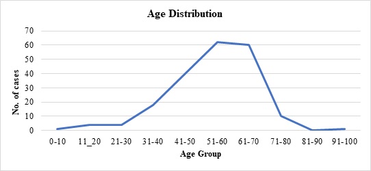
Results: 1) Malignant ascitic fluid cases are more common in older age group of patients and females outnumber the male patients substantially. 2) Adenocarcinoma is the most common histological type. 3) Ovary is the most common primary site of malignant peritoneal effusion. 4) Cytoblock preparation and immunohistochemistry on cytoblock can be very helpful in finding the primary site of origin in those cases where patients first presented to the hospital with signs of malignant ascites and no symptomatology related to primary site.
Conclusion: Cytological examination of serous effusions is an accurate, prompt, affordable technique having diagnostic and therapeutic implications. With the help of ancillary methods, the phenotype of cells can be identified, classify as well as confirm our diagnosis.
Downloads
References
Garrison RN, Kaelin LD, Galloway RH, Heuser LS. Malignant ascites. Clinical and experimental observations. Ann Surg. 1986;203(6):644-651. doi: https://doi.org/10.1097/00000658-198606000-00009.
Ayantunde AA, Parsons SL. Pattern and prognostic factors in patients with malignant ascites: a retrospective study. Ann Oncol. 2007;18(5):945-949. doi: https://doi.org/10.1093/annonc/mdl499.
Runyon BA, Montano AA, Akriviadis EA, Antillon MR, Irving MA, McHutchison JG. The serum-ascites albumin gradient is superior to the exudate-transudate concept in the differential diagnosis of ascites. Ann Intern Med. 1992;117(3):215-220. doi: https://doi.org/10.7326/0003-4819-117-3-215.
Becker G, Galandi D, Blum HE. Malignant ascites: systematic review and guideline for treatment. Eur J Cancer. 2006;42(5):589-597. doi: https://doi.org/10.1016/j.ejca.2005.11.018.
Kumar SK, Verma R, Sachan P, Bahl D. Malignant Ascites of Unknown Primary Tumour Site: A Clinical Dilemma. Int J Surg. 2006;11(1).
Udasimath S, Arakeril SU, Karigowdar MH, Yelikar BR. The Role of the Cell Block Method in the Diagnosis of Malignant Ascitic Fluid Effusions. J Clinic Diagnos Res. 2012;6(7):1280-1283.
Kushwaha R, Shashikala P, Hiremath S, Basavaraj HG. The cells in the pleural fluid and their value in the differential diagnosis. J Cytol 2008;25(4):138-143.
Dekker A, Bupp PA. The cytology of serous effusions. An investigation into the usefulness of cell blocks versus smears. Am J Clin Pathol 1978;70(6):855-860. doi: https://doi.org/10.1093/ajcp/70.6.855.
Turnage RH, Li DLB, McDonald JC. Abdominal wall umbilicus, peritoneum, mesenteries, omentum. In: Sabiston Textbook of Surgery – The biological basis of modern surgical practice. 17th ed. Philadelphia: Elsevier Saunders; 2004: 1182-1186.
Gia Khanh Nguyen. Essentials of fluid cytology. 2009:9-71
Bonito LD, Falconieri G, Colautti I, Bonifacio D, Dudine S. The positive pleural effusion. A retrospective study of the cytopathologic diagnosis along with the autopsy conformation. Acta Cytol. 1992; 36(3):329-32.
Abadi MA, Zakowski MF. Cytologic features of sarcomas in fluids. Cancer Cytopathol. 1998;84(2):71-76. doi: https://doi.org/10.1002/(sici)1097-0142(19980425)84:2%3C71::aid-cncr1%3E3.0.co;2-g.
Malik I, Abubakar S, Rizwana I, Alam F, Rizvi J, Khan A. Clinical features and management of malignant ascites. J Pak Med Assoc. 1991;41(2):38-40.
Parsons SL, Watson SA, Steele RJ. Malignant ascites. Br J Surg. 1996;83(1):6-14. doi: https://doi.org/10.1002/bjs.1800830104.
Elisabeth Smolle, Valentin Taucher, and Johannes Haybaeck Review: Malignant Ascites in Ovarian Cancer and the Role of Targeted Therapeutics. Anticancer Res. 2014;34(4)1553-1561.
Sereno M, Rodríguez-Esteban I, Gómez-Raposo C, et al. Lung cancer and peritoneal carcinomatosis. Oncol Lett. 2013;6(3):705-708. doi: https://dx.doi.org/10.3892%2Fol.2013.1468.
Kichian K, Bain VG. Jaundice, ascites and encephalopathy. In: Hanks GWC, Cherny NI, Calman K, Doyle D. (eds) Oxford Textbook of Palliative Medicine: 3rd edition. Oxford. Oxford University Press. 2005. p.507-519.
Copyright (c) 2020 Author (s). Published by Siddharth Health Research and Social Welfare Society

This work is licensed under a Creative Commons Attribution 4.0 International License.

 OAI - Open Archives Initiative