
Role of p16, Ki67 and CK17 in differentiating benign lesions, cervical intraepithelial neoplasia(CIN) and atypical immature squamous metaplasia(AIM) of uterine cervix
Abstract
Cervical intraepithelial neoplasia (CIN) is a premalignant lesion capable of progressing to cervical cancer. Despite the existing well-defined criteria, the histopathological diagnosis is subject to high rates of discordance among pathologists.
Aim: To study the role of p16, Ki67 and CK17 in differentiating benign lesions, cervical intraepithelial lesions(CIN) and atypical immature squamous metaplasia (AIM)and to improve intra and interobserver reproducibility of diagnosis of cervical neoplasia.
Material and Methods: In a cross sectional study, a total of 75 cervical biopsies including benign lesions (n=24), AIM (n=28), CIN (n=23) were studied and analyzed immunohistochemically using p16, Ki67 and CK17 immunomarkers. Data was evaluated using chi-square test.
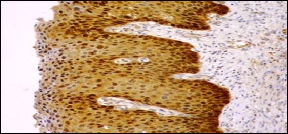
Results: p16 and Ki 67positivity were observed in 91.3% and 78.26% of CIN and 28.57% of AIM respectively. None of the benign lesions expressed p16 and Ki67while CK17 positivity was observed in 46.42% of CIN and 100% of AIM with 12.5% of benign lesions.
Conclusion: The three biomarkers (p16, CK17 and Ki67) had a high degree of sensitivity and specificity and appear to be a useful and reliable diagnostic adjunct to improve the routine diagnosis and reduce interobserver variability in cervical biopsy specimens. Immunohistochemical markers such as p16 alone or with Ki67 represents important tool for the pathologists in distinguishing high grade cervical dysplasia from its benign mimics such as AIM and reactive inflammatory lesion thus avoiding overtreatment.
Downloads
References
2. Fahey MT, Irwig L, Macaskill P. Meta-analysis of Pap test accuracy. Am J Epidemiol.1995Apr 1; 141(7s):680-9.
3. Sherman ME, Schiffman MH, Lorinez AT, Manos MM, Scott DR, Kuman RJ, et al. Toward objective Quality assurance in cervical cytopathology: Correlation of cytopathologic diagnosis with detection of high risk human papillomavirus types. Am J Clin Pathol.1994Aug; 102(2):182-7.
4. Singh N. HPV and Cervical cancer – prospectus for prevention through vaccination. Ind J Med Paedia Oncol.2005; 26(1):20-3.
5. Rosai J. Female reproductive system. In: Houston M, editor. Rosai and Ackerman’s surgical pathology. 9th Ed. New York: Mosby; 2004. pp. 1523–68.
6. Nam EJ, Kim JW, Hong JW, Jang HS, Lee SY, Jang SY, et al. Expression of the p16 and Ki-67 in relation to the grade of cervical intraepithelial neoplasia and high-risk human papillomavirus infection. J Gynecol Oncol. 2008Sep; 19(3):162–8.
7. Reuschenbach M, Seiz M, von KnebelDoeberitzC, Vinokurova S, Duwe A, Ridder R, et al. Evaluation of cervical cone biopsies for coexpression of p16INK4a and Ki-67 in epithelial cells. Int J Cancer. 2012Jan 15;130(2):388–94.
8. Keating JT, Cviko A, Riethdorf S, Riethdorf L, Quade BJ, Sun D, et al. Ki-67, cyclin E, and p16INK4 are complimentary surrogate biomarkers for human papilloma virus-related cervical neoplasia. Am J Surg Pathol. 2001Jul; 25(7):884–91. [PubMed]
9. Klaes R, Benner A, Friedrich T, Ridder R, Herrington S, Jenkins D, et al. p16INK4a immunohistochemistry improves interobserver agreement in the diagnosis of cervical intraepithelial neoplasia. Am J Surg Pathol. 2002Nov; 26(11):1389–99.
10. Galgano MT, Castle PE, Atkins KA, Brix WK, Nassau SR, Stoler MH. Using biomarkers as objective standards in the diagnosis of cervical biopsies. Am J Surg Pathol. 2010Aug; 34(8):1077–87.
11. Crum CP,Egawa K, Fu YS et al. Atypical immature metaplasia(AIM). A subset of human papillomavirus infection of the cervix. Cancer 1983;51:2214-2219.
12. Wentzensen N, von Knebel Doeberitz M. Biomarkers in cervical cancer screening. Dis Markers.2007; 23:315-30.
13. Eleutério J Jr, Giraldo PC, Gonçalves AK, Cavalcante DI, de Almeida Ferreira FV, Mesquita SM, et al. Prognostic markers of high-grade squamous intraepithelial lesions: The role of p16INK4a and high-risk human papillomavirus. Acta Obstet Gynecol Scand 2007; 86(1):94-8.
14. Cavalcante DM, Linhares LM, Pompeu MML, Givaldo PC, Eleutério J.The utility of p16 INK4a and Ki-67 to identify high-grade squamous intraepithelial lesion in adolescents and young women.Ind J Pathol Microbiol 2012;vol55(3):339-342.
15. Iaconis L, Hyjek E, Ellenson LH, Pirog E. p16 and Ki-67 immunostaining in atypical immature squamous metaplasia of the uterine cervix. Correlation with Humam papillomavirus detection. Arch Pathol Lab Med 2007Sep;131(9):1343-49.
16. Malpica A, Robboy SJ. Cervical benign and non neoplastic conditions. In: Robboy Js, Mutter GL, Prat J, Bentley RC, Russel P, Macolm C. Robboy’s pathology of the female Reproductive Tract. 2nd ed. London: Churchill livingstone; 2009.p.141-72.
17. Kalof AN, Evans FM, Simmons-Arnold L, Beatty BG, Cooper K. p16 ink4a immunoexpression and HPV in situ hybridization signal patterns:potential markers of high grade cervical intraepithelial neoplasia. AM J Surg Pathol. 2005May; 29(5):674-9.
18. Agoff SN, Lin P,Morihara J,Mao C, Kiviat N, Koustky L.p16ink4aexpression correlates with the degree of neoplasia: A comparison with ki67 expression and detection of high risk types. Mod Pathol. 2003Jul; 16(7):665-73.
19. Benevolo M, Mottolese M, Marandino F, Vocaturo G, Sindico R, Piperno G et al. iimmunohistochemical expression of p16 ink4a is predictive of HR-HPV infection in cervical low grade lesions. Mod Pathol. 2006 Mar; 19(3):384-91.
20. Focchi GR, Silva ID, Nogueira-de-Souza NC, Dobo C, Oshima CT,Stavale JN. Immunohistochemical exprerssion of p16ink4a in normal uterine cervix, nonneoplastic epithelial lesions and low grade squamous intraepithelial lesions. J Low Genit Tract Dis .2007Apr; 11(2):98-104.
22. Walts AE, Bose S. P16/ki67 immunostaining is useful in stratification of atypical epithelium of the cervix. Clin Med Pathol 2008Mar; 1:35-42.
23. Aslani FS, Safaei A, Pourjabali M , Momtahan M.Evaluation of ki67,p16 and ck17 markers in differentiating cervical intraepithelial neoplasia and benign lesions. Iran J Med Sci.2013Mar;38(1): 15-21.
24. Kruse AJ, Baak JP, de Bruin PC, Jiwa M, Snijders WP, Boodt PJ. Ki 67 immunoquantitation in cervical intraepithelial neoplasia (CIN): A sensitive marker for grading. J Pathol.2001 Jan; 193(1): 48-54.
25. Al-Saleh W, Delvenne P, Greimers R, Fridman V, Doyen J, Boniver J. Assesment of ki67 antigen immunostaining squamous intraepithelial lesions of the uterine cervix. Correlation with the histologic grade and human papillomavirus type.Am J Clin Pathol 1995Aug;104(2):154-60.
26. Ma L, Fisk JM, Zhang RR, Ulukus EC, Crum CP. Zheng W. Eosinophilic dysplasia of the cervix: a newly recognized variant of cervical squamous intraepithelial neoplasia. Am J Surg Pathol. 2004Nov; 28(11):1474-84. [PubMed]

 OAI - Open Archives Initiative