
Clinicopathological study of Meningioma
Abstract
Introduction: Meningiomas are tumors that arise from the meningothelial cells. They are commonly located at intracranial, intraspinal or occasionally ectopic site. They show histological diversity and are categorized into three grades. This grading helps in predicting their behaviour and deciding treatment strategy.
Aims and Objectives: To study the incidence, anatomical location, sex and age Predilection, histological variants and grading of meningiomas based on WHO 2016 classification. To correlate clinical features and radiological findings with those of histopathological findings.
Materials and Methods: The study is carried out in the Department of Pathology, Dhiraj General Hospital, Piparia from November 2016 to July 2018. 30 tumors specimen diagnosed as meningioma by radiology and neurosurgery department, sent to department of pathology were included in the study. Analysis of histological features, typing and grading of all cases was done according to WHO 2016 classification of meningioma.
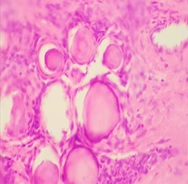
Results: Total 30 meningioma tumors were included in the study. Most of them were intracranial, predominantly involving the posterior fossa of brain, females and the 41-60 age group. The most common histological subtype was psammomatous followed by meningothelial. Majority (93.33%) were benign grade I tumors. In 90% cases radiological diagnosis matched exactly with histopathological diagnosis.
Conclusion: Meningiomas are slow growing tumors arising from the meningothelial cells accounting for 15-30 % of all CNS neoplasms showing a variety of histological patterns, more common in women, predominantly Grade I tumors. Recurrence of tumors depends on histological grade and extent of surgery.
Downloads
References
Perry a, Louis DN, Scheithauer BW, Budka H, von Deimling A: Meningiomas in WHO Classification of Tumours of the Central Nervous System, 4th Edition, IARC press, Lyon 2007;1:164-172.
Lamszus K. Meningioma pathology, genetics, and biology. J Neuropathol Exp Neurol. 2004;63(4):275–286. doi: https://doi.org/10.1093/jnen/63.4.275.
Gattu V. Histopathological analysis of meningiomas- A retrospective study. SAS J Surg. 2017;3(1):25-29.
Louis DN, Ohgaki H, Wiestler OD, Cavenee WK, Burger PC, Jouvet A, et al Acta Neuropathol. Acta Neuropathol. 2007;114(2):97-109. Epub 2007 Jul 6. doi: https://doi.org/10.1007/s00401-007-0243-4.
Perry A, Stafford SL, Scheithauer BW, Suman VJ, Lohse CM. Meningioma grading: an analysis of histologic parameters. Am J Surg Pathol. 1997;21(12):1455-1465.
Miller DC. Meningiomas. In: Miller DC, editor. Modern surgical pathology. Cambridge University Press. 2009. pp. 217–236.
Willis J, Smith C, Ironside JW, Erridge S, Whittle IR, Everington D. The accuracy of meningioma grading: a 10-year retrospective audit. Neuropathol Appl Neurobiol. 2005;31(5):141-149. doi: https://doi.org/10.1111/j.1365-2990.2004.00621.x.
Commins DL, Atkinson RD, Burnett ME. Review of meningioma histopathology. Neurosurg Focus. 2007;23(4):E3. doi: https://doi.org/10.3171/FOC-07/10/E3.
Louis DN, Perry A, Reifenberger G, von Diemling A, Figarella-Branger D, Cavenee WK, et al. Acta Neuropathol. 2016;131(6):803-820. doi: https://doi.org/10.1007/s00401-016-1545-1. Epub 2016 May 9.
Rogers L, Gilbert M, Vogelbaum MA. Intracranial meningiomas of atypical (WHO grade II) histology. J Neurooncol. 2010;99(3):393–405. doi: https://doi.org/10.1007/s11060-010-0343-1.
Deborah L. Commins S, Roscoe D. Atkinson Margaret E. Burnett, Review of meningioma histopathology; Neurosurg Focus 2007;23(4):E3. doi: https://doi.org/10.3171/FOC-07/10/E3.
Reddy R. Histopathological spectrum of meningioma and its variants. Asian Pac J Health Sci. 2016;3(1):151-155.
Wang DJ, Zheng MZ, Gong Y, Xie Q, Wang Y, Cheng HX, et al. Papillary meningioma: clinical and histopathological observations. Int J Clinic Experiment Pathol. 2013;6(5):878-888.
Juong lee, Meningiomas; Diagnosis and treatment and outcome of meningiomas; 8th Ed, 2008.
Germano IM, Michael S. Edwards B, Davis RL, Schiffer D. Intracranial meningiomas of the first two decades of life. J Neurosurg. 1994;80(3):447-453. doi: https://doi.org/10.3171/jns.1994.80.3.0447
Mehta N, Bhagwati S, Parulekar G. Meningiomas in children: A study of 18 Cases. J Pediatric Neurosci. 2009;4(2):61-65. doi: https://doi.org/10.4103/1817-1745.57322.
Yoon SH, Chung CK, Jahng TA. Surgical Outcome of Spinal Canal Meningiomas. J Korean Neurosurg Soc. 2007;42(4):300-304. doi: https://doi.org/10.3340/jkns.2007.42.4.300.
Gottfried ON, Gluf W, Hinojosa AQ, Kan P, Schmidt MH. Spinal meningiomas: Surgical management and outcome Neurosurg Focus 2003;14(6):e2. doi: https://doi.org/10.3171/foc.2003.14.6.2.
Elizabeth B, Clauis MD, Melissa L, Bondy MD, Black PM. Epidemiology of Intra cranial meningiomas. Neurosurg. 2005;57(6):1088-1095. doi: https://doi.org/10.1227/01.NEU.0000188281.91351.B9
Longstreth WT. Dental X rays and the risk of intracranial meningiomas. Cancer. 2004;100(5):1026-1034. doi: https://doi.org/10.1002/cncr.20036
Bi WL, Abedalthagafi M, Horowitz P. Agarwalla PK, Mei Y, Aizer AA et al. Genomic landscape of intracranial meningiomas. J Neurosurg. 2016; 125(3):525-535. doi: https://doi.org/10.3171/2015.6.JNS15591.
Perry A, Gutmann DH, Reifenberger G. Molecular pathogenesis of meningiomas. J Neuro Oncol. 2004; 70(2):183-202.
Engelhard HH. Progress in the diagnosis and treatment of patients with meningiomas. Part I: diagnostic imaging, preoperative embolization. Surg Neurol. 2001;55(2):89-101.
Grondahl TB, Moen BH, Torp SH. The histopathological spectrum of human meningiomas. Int J Clin Exp Pathol. 2012;5(3):231-242.
Perry A, Scheithauer BW, Stafford SL. “Malignancy” in Meningiomas A clinicopathological Study of 116 patient with Grading Implications. Cancer. 1999;85(9):2046-2056.
Violaris K, Katsarides V, Sakellariou P; The recurrence rate in meningiomas: Analysis of tumor location,histological grading, and extent of resection; Open J Mod Neurosurg. 2012:2(1):6-10. doi: https://doi.org/http://dx.doi.org/10.4236/ojmn.2012.21002.
Adlakha A, Rao K, Adlakha H. Meningioma metastatic to the lung. Mayo Clin Proc. 1999;74:1129-1133.
Takei H, Powell SZ. Tumor-to-tumor metastasis to the central nervous system. Neuropathol. 2009;29(3):303-308. doi: https://doi.org/10.1111/j.1440-1789.2008.00952.x.
Lakshmi SS. Meningiomas: A Clinicopathological study. Int J Med Res Heal Sci. 2015;4(4):827-831. doi: https://doi.org/10.5958/2319-5886.2015.00164.2.
Fonkem E, Dandashi JA, Stroberg E, Garrett D, Harris FS, Nihum IM, et al. A retrospective analysis of meningioma in Central Texas. J Epidemiol Glob Health. 2016;6(2):87-93. doi: https://dx.doi.org/10.1016/j.jegh.2016.01.001.
Varlotto J, Flickinger J, Pavelic MT. Distinguishing grade I meningioma from higher grade meningiomas without biopsy. Oncotarget.2015;6(35):38421-38428. doi: https://doi.org/http://doi.org/10.18632/oncotarget.5376.
Raza AKMM, Ahmed F, Munni TA. Histomorphological spectrum of meningioma with variants and grading. Adv Surg Res. 2017;1(1):15-17.
Shah S, Gonsai RN, Makwana R. Histopathological study of meningioma in civil hospital Ahmedabad. Int J Cur Res Rev. 2013;5(3):76-82. doi: https://doi.org/10.5958/2395-1184.2019.00003.2.
Joseph, Wanjeri et al. Histology and clinical pattern of meningiomas at the Kenyatta National Hospital Nairobi, Kenya. A thesis submitted for the award of the degree of master of medicine in neurosurgery. University of Nairobi; 2011.
Nath HD, Mainuddin MD, Ehsammahmood KU. Surgical outcome of supratentorial meningioma. A study of 25 cases. JCMCTA 2009;20(1):41-44. doi: https://doi.org/10.3329/jcmcta.v20i1.4934.
Gadgil NM, Margam SR, Chaudhuri CS, Kumavat PV. The histopathological spectrum of meningeal neoplasms. Indian J Pathol Oncol. 2016;3(3):432-436. doi: https://doi.org/10.5958/2394-6792.2016.00081.8.
Patil PR, Sondankar D. Clinicopathological Study of Meningioma. Int J Med Res Rev. 2016;4(4):592-601. doi: https://doi.org/10.17511/ijmrr.2016.i04.20.
Jat KC, Vyas SP, Bihari NA, Mehra K. Central nervous system tumors: a histopathological study. Int J Res Med Sci. 2016;4(5):1539-1545. doi: http://dx.doi.org/10.18203/2320-6012.ijrms20161225.
Narmadha R, Dhanalakshmi S, Priyadharshini M. Histomorphological spectrum of central nervous system tumours- A three-year retrospective descriptive study in atertiary care centre. J Evolution Med. Dent Sci. 2017;6(43):3362-3366. doi: https://doi.org/10.14260/jemds/2017/728.
Samadi N, Ahmadi SA. Meningioma: A clinicopathological evaluation. Malays J Med Sci. 2007;14(1):46-52.
Babu S, Uppin SG, Uppin MS. Meningiomas: Correlation of Ki67 with histological grade. Neurol India. 2011;59(2):204-207. doi: https://doi.org/10.4103/0028-3886.79140.
Prayson R.A. (2009) Pathology of Meningiomas. In: Lee J.H. (eds) Meningiomas. Springer, London.
Taghipour M, Rakei SM, Monabati A. The role of estrogen and progesterone receptors in grading of the malignancy of meningioma. Iran Red Crescent Med J.2007;9(1):17-21.
Copyright (c) 2020 Author (s). Published by Siddharth Health Research and Social Welfare Society

This work is licensed under a Creative Commons Attribution 4.0 International License.

 OAI - Open Archives Initiative