
Evaluating the spectrum of histomorphological patterns on endoscopic biopsy in patients with upper gastrointestinal tract disorders
Abstract
Introduction: The upper gastrointestinal tract (UGT) disorders are more common complaints in clinical practice and have got high degree of mortality and morbidity. Many different types of lesions can affect the upper gastrointestinal tract and can be classified as congenital anomalies, infections, inflammation and neoplastic lesions.
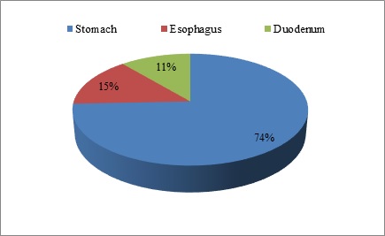
Material and Methods: A total of 152 cases of upper gastrointestinal tract biopsies are included in this study, out of which 113 cases were gastric biopsies, 22 cases were esophageal biopsies and the remaining 17 cases were duodenal biopsies. Present study was carried out in the Department of Pathology at Sri Venkateshwaraa Medical College Hospital and Research Centre, Puducherry for a period of one year between March 2018 and February 2019. All the biopsies were performed using fiberoptic endoscopy. Endoscopic biopsy in combination with histopathological examination plays an important role in the early diagnosis of determining UGT lesions. Also, special stains like Giemsa, Warthin starry stain was used to demonstrate Helicobacter pylori. Aim of the study is to evaluate the histomorphological patterns of upper gastrointestinal tract disorders on endoscopic biopsy and to correlate various upper gastrointestinal tract disorders in correspondence to clinical parameters.
Results: It is reported among 152 cases of UGT biopsies 137 were non-neoplastic lesions and 15 were neoplastic. Commonly affected age group was 31-40 years followed by 41 to 50 years. As per the present study, males were affected more predominantly than females. Out of the 22 cases from esophageal biopsies, 16 cases showed non-neoplastic lesions and 6 were neoplastic. The most common non-neoplastic lesion was chronic nonspecific esophagitis and neoplastic lesion reported was squamous cell carcinoma. Among 113 gastric biopsies, 104 cases were non-neoplastic lesions and 9 were neoplastic lesions. Adenocarcinoma was the predominant neoplastic lesion of stomach. All the 17 duodenal biopsies showed non -neoplastic lesions.
Downloads
References
Blackstone MO. Endoscopic interpretation. Normal and pathologic appearances of Gastrointestinal tract. New York: Raven Press; 1984. P 13-15.
Gulia SP, Chaudhury M, Noorunnisa N, Balakrishnan CD, Balagurunathan K. Interpretation of Upper GastroIntestinal Tract Endoscopic Mucosal Biopsies–A Study Conducted In Teaching Hospital In Puducherry, India. Int J Med Health Sci. 2012;1(3):17-24.
Afzal S, Ahmad M, Mubarik A, Saeed F, Rafi S, Saleem N, Qur AH. Morphological spectrum of gastric lesions-Endoscopic biopsy findings. Pak Armed Forces Med J. 2006;56(2):143-149.
Memon F, Baloch K, Memon AA. Upper gastrointestinal endoscopic biopsy; Morphological spectrum of lesions. Professional Med J. 2015;22(12):1574-1579. doi: https://doi.org/10.17957/TPMJ/15.3027.
Hirachand S, Sthapit RR, Gurung P, Pradhanang S, Thapa R, Sedhai M, RegmiS.Histopathological spectrum of upper gastrointestinal endoscopic biopsies. J BP Koirala Inst Health Sci. 2018;1(1):67-74. doi: https://doi.org/10.3126/jbpkihs.v1i1.19760.
Krishnappa R, Horakerappa MS, Mangala AK, Gouri M. A study onhistopathologic spectrum of upper gastrointestinal tract endoscopic biopsies. Int J Medical Res Health Sci. 2013;2(3):418-424. doi: https://doi.org/10.5958/j.2319-5886.2.3.073.
Teriaky A, Al Nasser A, McLean C, Gregor J, Yan B. The utility of endoscopic biopsies in patients with normal upper endoscopy. Can J Gastroenterol Hepatol. 2016;2016. doi: http://dx.doi.org/10.1155/2016/3026563.
Saha. Studies on Helicobacter pylori: National Institute of Cholera and Enteric Diseases. Annual Report 2004-2005.
Singh V, Trikha B, Vaiphei K, Nain CK, Thennarasu K, Singh K. Helicobacter py¬lori: Evidence for spouse-to-spouse transmission. J Gastroenterol Hepatol. 1999;14(6):519-522. doi: https://doi.org/10.1046/j.1440-1746.1999.01908.x.
Kobayashi O, Eishi Y, Ohkusa T, Ishige. Gastric mucosal density of H. Pylori estimated by real-time PCR compared with results of urea breath test and histological grading. J Med Microbiol. 2002;51(4):305-311. doi: https://doi.org/10.1099/0022-1317-51-4-305.
Shennak MM, Tarawneh MS, Al Sheik. Upper gastrointestinal diseases in symptomatic Jordanians: A prospective study. Ann Saudi Med. 1997;17(4):471-474. doi: https://doi.org/10.5144/0256-4947.1997.471.
National Cancer Registry Programme. First All India Report 2001-2002. Vol 1. Indian Council of Medical Research. Bangalore, India. 2004.
Nafees A Qureshi, Michael T Hallissey, John W, Fielding Outcome of indexupper gastrointestinal endoscopy in patients presenting with dysphagia in atertiary care hospital - A 10 years review. BMC Gastroenterol. 2007;7:43. doi: https://doi.org/10.1186/1471-230X-7-43.
International Agency for Research on Cancer. Latest world cancer statistics Global cancer burden rises to 14.1million new cases in 2012: Marked increase in breast cancers must be addressed. World Health Organization. 2013;12. Available at https://www.iarc.fr/wp-content/uploads/2018/07/pr223_E.pdf.
Shin A, Won YJ, Jung HK, Kong HJ, Jung KW, Oh CM, et al. Trends in incidence and survival of esophageal cancer in Korea: Analysis of the Korea Central Cancer Registry Database. J Gastroenterol Hepatol. 2018;33(12):1961-1968. doi: https://doi.org/10.1111/jgh.14289.
Khandelia R, Saikia M. Histopathologic Spectrum of Upper Gastrointestinal Tract Mucosal Biopsies: A Prospective Study. Int J Med Sci Clinic Invent. 2017;4(11):3314-3316. doi: https://doi.org/10.18535/ijmsci/v4i11.11.
Kadish SL, Kochman ML.Endoscopic diagnosis and management of gastrointestinal malignancy. Oncol. 1995;9(10):967-983.
Laine L, Lewin DN, Naritoku W, Cohen H. Prospective comparison of H&E, Giemsa, and Genta stains for the diagnosis of Helicobacter pylori. Gastrointest endos. 1997;45(6):463-467. doi: https://doi.org/10.1016/S0016-5107(97)70174-3.
Fallone CA, Loo VG, Lough J, Barkun AN. Hematoxylin and eosin staining of gastric tissue for the detection of Helicobacter pylori. Helicobacter. 1997;2(1):32-35. doi: https://doi.org/10.1111/j.1523-5378.1997.tb00054.x
Pandya HB, Agravat HH, Patel JS, Sodagar NR. Emerging antimicrobial resistance pattern of H. Pylori in central Gujarat. Indian J Med Microbiol. 2014;32(4):408-413. doi: https://doi.org/10.4103/0255-0857.142256.
Sgambato D, Visciola G, Ferrante E, Miranda A, Romano L, Tuccillo C, et al. Prevalence of H. Pylori infection in sexual partners of H. pylori-infected subjects: Role of gastroesophageal reflux. United Europe Gastroenterol J. 2018;6(10):1470-1476. doi: https://doi.org/10.1177/2050640618800628.
Morilla A, Melón S, Álvarez-Argüelles ME, Armesto E, Villar H, de Oña M. Utility of normalized genome quantification of H. Pylori in gastric mucosa using an in-house real-time polymerase chain reaction. PloS one. 2017;12(6):e0178674. doi: https://doi.org/10.1371/journal.pone.0178674.
Ozturk S, Serinsoz E, Kuzu I, Ensari A, Erden E, Kansu A, et al. The Sydney System in the assessment of gastritis: Inter-observer agreement. The Turkish J Gastroenterol. 2001;12(1): 36-39.
Hassan TM, Al-Najjar SI, Al-Zahrani IH, Alanazi FI, Alotibi MG. H. Pylori chronic gastritis updated Sydney grading in relation to endoscopic findings and H. pylori IgG antibody: diagnostic methods. J Microscop Ultrastruct. 2016;4(4):167-174. doi: https://doi.org/10.1016/j.jmau.2016.03.004.
Sheikh BA, Hamdani SM, Malik R. Histopathological spectrum of lesions of upper Gastrointestinal tract-A study of endoscopic biopsies. GJMEDPH.2015;4(4):1-8.
Khatib WM, Demde RB, Aher VC, Patel PM. Histopathological Spectrum of Non-Malignant Lesions of Gastrointestinal Tract-An Institutional Stud. IOSR J Dent Med Sci. 2016;15(10):113-116
Shepherd NA, Valori RM. The effective use of gastrointestinal histopathology:guidance for endoscopic biopsy in the gastrointestinal tract. Front Gastroenterol. 2014;5(2):84-7. doi: http://dx.doi.org/10.1136/flgastro-2013-100413.
Shennak MM, Tarawneh MS, Al Sheikh TM, Pattern of upper gastrointestinal disease in symptomatic Jordanians: a prospective endoscopic study of 5657 patients, Dirasat. 1998;25(2):69-81.
Islam SM, Ahmed AM, Ahmad MS, Hafiz SA. Endoscopic and histologicDiagnosis of upper gastrointestinal lesions, Experience in a port city of Bangladesh. Chattagram Maa-O-Shishu Hospital Med College J. 2014;13(3):11-14. doi: https://doi.org/10.3329/cmoshmcj.v13i3.20997.
Nwafor CC, Nwafor NN, Etuk EB, Kanu O. Histopathological spectrum of gastrointestinal lesions seen in university of uyo teaching hospital, South–South Nigeria. Ann Trop Pathol. 2019;10(1):27-33. doi: https://doi.org/10.4103/atp.atp_40_18.
Khandelia R, Saikia M. Histopathologic Spectrum of Upper GastrointestinalTract Mucosal Biopsies: A Prospective Study. Int J Med Sci Clinic Invent. 2017;4(11):3314-3316. doi: https://doi.org/10.18535/ijmsci/v4i11.11.
Hussain SI, Reshi R, Akhter G, Beigh A. Clinico histopathological study ofupper gastrointestinal tract endoscopic biopsies. Int J Curr Res Rev. 2015;7(16):78-85.
Thapa R, Lakhey M, Yadav PK, Kandel P, Aryal C, Subba K. Histopathologicalstudy of endoscopic biopsies. J Nepal Med Assoc.2013;52(190):354-356. doi: https://doi.org/10.31729/jnma.2116.
Prasaad PR, Rao B. Histopathological spectrum of gastrointestinal lesions-anexperience in a tertiary care centre in South India. Int J Res Med Sci. 2016;4(8):3407-3412. doi: http://dx.doi.org/10.18203/2320-6012.ijrms20162302.
Copyright (c) 2020 Author (s). Published by Siddharth Health Research and Social Welfare Society

This work is licensed under a Creative Commons Attribution 4.0 International License.

 OAI - Open Archives Initiative