
Tumoral calcinosis simulating sarcoma
Abstract
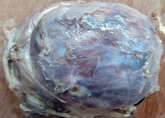
Tumoral calcinosis is a rare clinical and pathological entity, characterized by deposition of calcium in different sites of human body particularly in peri-articular tissues. It incidence is usually seen in young adults as a slowly growing, painless, firm to bony hard mass. Asymptomatic when small in size but larger masses can cause pressure symptoms due to compression of surrounding tissues or restriction of mobility of the joints. It can be predicted by typical radiological findings with the help of serum calcium and phosphorus levels but is confirmed only by histopathological examination. Many times, it isn’t very easy to diagnose this condition preoperatively and can be mistaken as soft tissue sarcoma both clinically and radiologically. Subtyping of this lesion depends on serum calcium, phosphorus levels, family history and co-morbid conditions like chronic renal failure. Hemodialysis, hypervitaminosis-D, milk-alkali syndrome, hyperparathyroidism, etc are responsible for the secondary causes. Treatment modality depends on the subtype, which can be medical or by surgical intervention. The present study was a case of adolescent male with a slowly growing swelling over the right hip region for 4 years. There was no traumatic history or any significant family history. Serum calcium and phosphorus levels were within normal limits. Radiology showed well-demarcated lobulated solid-cystic mass swelling measuring 7.8x3.9x3.5cm suggestive of sarcoma. The tumor was excised and sent for histopathological examination. Grossly, it was a ovoid solid-cystic firm to hard mass, microscopy showed large areas of dystrophic calcification with cystic spaces surrounded by foreign body giant cell reaction. Correlating with history, clinical findings, biochemical findings, the diagnosis of primary normophosphatemic tumoral calcinosis was made.
Downloads
References
McClatchie S, Bremner AD. Tumoral calcinosis--an unrecognized disease. Br Med J. 1969;1:153-155. doi: https://doi.org/10.1136/bmj.1.5637.142-a.
Smack D, Norton SA, Fitzpatrick JE. Proposal for a pathogenesis-based classification of tumoral calcinosis. Int J Dermatol. 1996;35(4):265-271. doi: https://doi.org/10.1111/j.1365-4362.1996.tb02999.x.
Hershkovitz D, Gross Y, Nahum S, Yehezkel S, Sarig O, Uitto J et al. Functional characterization of SAMD9, a protein deficient in normophosphatemic familial tumoral calcinosis. J Invest Dermatol. 2011;131(3):662-669. doi: https://doi.org/10.1038/jid.2010.387.
Topaz O, Shurman DL, Bergman R, Indelman M, Ratajczak P, Mizrachi M et al. Mutations in GALNT3, encoding a protein involved in O-linked glycosylation, cause familial tumoral calcinosis. Nat Genet. 2004;36(6): 579-581. doi: https://doi.org/10.1038/ng1358.
Olsen KM, Chew FS. Tumoral calcinosis: pearls, polemics, and alternative possibilities. Radiograph. 2006; 26(3):871-885. doi: https://doi.org/10.1148/rg.263055099.
Inclan A, Leon PP, Camejo M. Tumoral calcinosis. J Am Med Ass. 1943;121:490-495. doi: https://doi.org/10.1001/jama.1943.02840070018.
Kannan S, Ravikumar L, Mahadevan S, Natarajan M, Satya A, Bhat R, et al. Tumoral calcinosis with Vitamin D deficiency. Saudi J Kidney Dis Transpl. 2008; 19(6): 960 963.
Mohamed S, Jong Hun J, Weon Yoo K. Tumoral calcinosis of the foot with unusual presentation in an 11 year old boy: A case report and review of literature. J Postgrad Med. 2007;53(4):247 249. doi: https://doi.org/10.4103/0022-3859.37513.
Shaukat YM, Malik EF, Al Rashid M, Cannon SR. Large tumoral calcinosisin the gluteal region: A case report. Ortop Traumatol Rehabil 2013;15(5):495 499. doi: https://doi.org/10.5604/15093492.1084363.
Lykoudis EG, Seretis K, Ristanis S. Huge recurrent tumoral calcinosis needing extensive excision and reconstruction: Report of a rare case and brief literature review. Aesthetic Plast Surg. 2012;36(5):1194 1197. doi: https://doi.org/10.1007/s00266-012-9923-0.
Chew FS, Roberts CC. Musculoskeletal imaging: a teaching file. 2nd ed. New York, NY: Lippincott Williams & Wilkins, 2006; 233.
Jones BC, Sundaram M, Kransdorf MJ. Synovial sarcoma: MR imaging findings in 34 patients. AJR Am J Roentgenol. 1993;161:827-830.
Kransdorf MJ, Meis JM. Extraskeletal osseous and cartilaginous tumors of the extremities. Radiograph. 1993;13(4):853-884. doi: https://doi.org/10.1148/radiographics.13.4.8356273.
Flemming DJ, Murphey MD, Shekitka KM, Temple HT, Jelinek JJ, Kransdorf MJ. Osseous involvement in calcific tendonitis: a retrospective review of 50 cases. AJR Am J Roentgenol. 2003;181(4):965-972. doi: https://doi.org/10.2214/ajr.181.4.1810965.
Lin YP, Chen CH. Huge tumoral calcinosis of the buttock. Formosan J Surg. 2014;47(1):23 27. doi: https://doi.org/10.1016/j.fjs.2013.06.010.
Liu X, Hong S, Chen G, Tu C. Large tumoral calcinosis and pathological femur fracture in a hemodialysis patient with secondary hyperparathyroidism. Int Surg J. 2015;2 (1):91 94. doi: https://doi.org/10.5455/2349-2902.isj20150219.
Copyright (c) 2020 Author (s). Published by Siddharth Health Research and Social Welfare Society

This work is licensed under a Creative Commons Attribution 4.0 International License.

 OAI - Open Archives Initiative