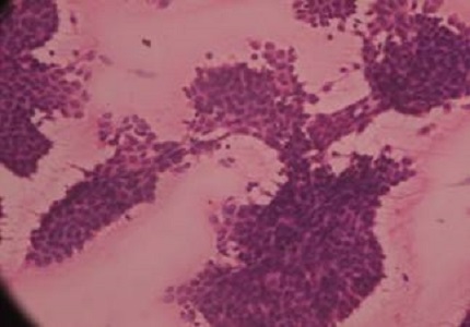
Correlative study of FNAC and histopathology for breast lesions
Abstract
Introduction: Fine needle aspiration cytology has become increasingly popular in diagnosis of palpable breast masses as it is sensitive, specific, expedient, economical and safe for screening of breast lesions. It has high sensitivity and specificity. The aim of the study was to categorize breast lesions and correlate the fine needle aspiration cytology diagnosis with histo-pathological findings.
Methods: A Two years study was carried out on female and male patients with age 10-72, who visited hospital with complaint of breast lump, pain in the breast or discharge from the nipple.
Results: Tumors on right breast is higher in percentage (54%) and 66% of tumors are less than 6 cm in size. Two cases, which found to be malignant with FNAC have become benign in biopsy test.
Conclusion: The current study initiated to find the effectiveness of this technique and compared with biopsy methods.
Downloads
References
2. Mulazim Hussain Bukhari, Madiha Arshad, Shahid Jamal, Shahida Niazi, Shahid Bashir, Irfan M. Bakhshi, and Shaharyar. Use of Fine-Needle Aspiration in the Evaluation of Breast Lumps. Patholog Res Int. 2011; 2011: 689521. [PubMed]
3. Rubin M, Horiuchi K, Joy N et al. Use of fine needle aspiration for solid breast lesions is accurate and cost-effective. Am J Surg. 1997 Dec;174(6):694-6; discussion 697-8. [PubMed]
4. Bhaskar Thakkar, Malay Parekh, N J Trivedi, A S Agnihotri, Uravashi Mangar. Role of fine needle aspiration cytology in palpable breast lesions and its correlation with histopathological diagnosis. National Journal of Medical Research . 2014;4(4):283-288.
5. Rahman MZ, Sikder AM, Nabi SR. Diagnosis of breast lump by fine needle aspiration cytology and mammography. Mymensingh Med J. 2011 Oct;20(4):658-64. [PubMed]
6. Neha Amrut Mahajan, C.P Bhale, S.S Mulay. Fine Needle Aspiration Cytology of Breast Lesions and Correlation with Histopathology; A 2 Year Study. International Journal of Health Sciences & Research. 2013;3(2);55-65.
7. Walid E. Khalbuss, M.D., Abiy Ambaye, Steve Goodison, Asif Loya, and Shahla Masood. Papillary Carcinoma of the Breast in a Male Patient With a Treated Prostatic Carcinoma Diagnosed by Fine-Needle Aspiration Biopsy: A Case Report and Review of the Literature. Diagn Cytopathol. 2006 Mar; 34(3): 214–217.
8. Ascoli V, Scalzo CC, Bruno C, Facciolo F, Lopergolo M, Granone P, Nardi F. Familial pleural malignant mesothelioma: clustering in three sisters and one cousin. Cancer Lett. 1998;130(1-2):203–207.
9. Azuma K, Uchiyama I, Chiba Y, Okumura J. Mesothelioma risk and environmental exposure to asbestos: past and future trends in Japan. Int J Occup Environ Health. 2009 Apr-Jun;15(2):166-72. [PubMed]
10. Bani-Hani KE, Gharaibeh KA. Malignant peritoneal mesothelioma. J Surg Oncol. 2005 Jul 1;91(1):17-25. [PubMed]
11. Baker PM, Clement PB, Young RH. Malignant peritoneal mesothelioma in women: a study of 75 cases with emphasis on their morphologic spectrum and differential diagnosis. Am J Clin Pathol. 2005;123(5):724–737.
12. Baumann F, Rougier Y, Ambrosi JP, Robineau BP. Pleural mesothelioma in New Caledonia: an acute environmental concern. Cancer Detect Prev. 2007;31(1):70-6. Epub 2007 Feb 23.
13. Berry G, Roger J, Pooley FD. Mesotheliomas. Asbestos exposure and lung burden. In: Bignon J, Peto J, Saracci R, editors. Non occupational exposure to mineral fibers. Lyon, France: 1989. pp. 486–496. IARC scientific publication No 90.
14. Hindle WH, Payne PA, Pan EY. The use of fine-needle aspiration in the evaluation of persistent palpable dominant breast masses. Am J Obstet Gynecol. 1993 Jun;168(6 Pt 1):1814-8; discussion 1818-9.
15. Lee HC, Ooi PJ, Poh WT, Wong CY. Impact of inadequate fine-needle aspiration cytology on outcome of patients with palpable breast lesions. Australian and New Zealand Journal of Surgery. 2000;70(9):656–659.
16. Khatun H, Tareak-Al-Nasir N, Enam S, Hussain M, Begum M. Correlation of fine needle aspiration cytology and its histopathology in diagnosis in breast lumpus. Bangladesh Med Res Counc Bull. 2002 Aug;28(2):77-81.
17. Nemenqani D, Yaqoob N. Fine needle aspiration cytology of inflammatory breast lesions. Journal of the Pakistan Medical Association. 2009;59(3):167–170.
18. 20. Das DK, Sodhani P, Kashyap V, Parkash S, Pant JN, Bhatnagar P. Inflammatory lesions of the breast: Diagnosis by fine needle aspiration. Cytopathology. 1992;3(5):281–289. [PubMed]
19. Bardales RH, Stanley MW. Benign spindle and inflammatory lesions of the breast: diagnosis by fine- needle aspiration. Diagnostic Cytopathology. 1995;12(2):126–130.
20. Guray M, Sahin AA. Benign breast diseases: classification, diagnosis, and management. Oncologist. 2006 May;11(5):435-49.
21. Lee KC, Chan JK, Gwi E. Tubular adenosis of the breast: a distinctive benign lesion mimicking invasive carcinoma. Am J Surg Pathol. 1996 Jan;20(1):46-54.


 OAI - Open Archives Initiative


