
Spectrum of various skin nodule mimicking cutaneous adnexal tumour
Abstract
Introduction: Cutaneous adnexal tumors are the group of uncommon tumour usually misdiagnosed clinically with various soft tissue tumour. Therefore, histopathology is the gold standard to diagnose their histogenesis. The aim of the present study is to find out the occurrence of skin adnexal tumors according to age, sex and site distribution along with histopathological diagnosis.
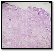
Materials and Methods: This retrospective study was conducted in Department of Pathology of Pacific Institute of Medical sciences, Udaipur from September 2016 to August 2019. The formalin fixed paraffin embedded tissue section was stained with hematoxylin and eosin stain for microscopical analysis.
Results: A total number of 41 adnexal tumors out of which 22 males and 19 females were studied out of which 36 cases were diagnosed as benign and 5 cases were reported as malignant. The most common cutaneous adnexal tumors diagnosed as sweat gland origin followed by hair follicle, sebaceous gland and pagets disease of nipple. Majority of the cutaneous adnexal tumors reported in head and neck site in a fourth to sixth decades of life.
Conclusion: Architectural features help us to diagnose different types of cutaneous adnexal tumors.
Downloads
References
Sahu A, Dilip KS, Nayak SK, Agrawal KC. Skin Adnexal Tumors: A Histopathological Study of 60 Cases at a Tertiary Care Centre. Ann Pathol Lab Med. 2018;5(3):215-220. doi: https://doi.org/10.21276/APALM.1787.
Valand AG, Ansari SAH, Sinha RK, Jadhav VC. A clinicopathological study of adnexal tumors of skin in a tertiary care research hospital. Int J Health Sci Res. 2016; 6(12):52-58.
Gandhi R, Srinivasan S. A Morphologic Study of Cutaneous Adnexal Tumours. Nat J Lab Med. 2016;5(4): 8-11. doi: https://doi.org/10.7860/NJLM/2016/21800:2161.
Prasad BVS, Faheem MK, Anuradha B, Lakshmi AV, Sreenivasulu M, Prasad DN. Histopathological Evaluation and Review of Cutaneous Adnexal Tumors (Cats) – A Research Study. IOSR J Dent Med Sci. 2018;17(2):7-11. doi: https://doi.org/10.9790/0853-1702030711.
Kaur R, Kumar V, Mehra K, Gupta N, Singh A. Histopathological evaluation of Skin Tumours. Indian J Pathol Oncol. 2016;3(4);627-631. doi: https://doi.org/10.5958/2394-6792.2016.00116.2.
Le Boit PE, Burg G, Weedon D, Sarasin A. World Health Organization Classification of Tumours Pathology and Genetics of Skin Tumours. 3rd Edition. France: IARC Press, International Agency for Research on Cancer, 150 cours Albert Thomas, F-69008 Lyon, France; 2006.
Fulton EH, Kaley JR, Gardner JM. Skin Adnexal Tumors in Plain Language A Practical Approach for the General Surgical Pathologist. Arch Pathol Lab Med. 2019;143(7):832-851. doi: https://doi.org/10.5858/arpa.2018-0189-RA.
Kaur K, Gupta K, Hemrajani D, Yadav A, Mangal K. Histopathological analysis of skin adnexal tumors: A three-year study of 110 cases at a tertiary care center. Indian J Dermatol 2017;62(4):400-406. doi: https://doi.org/10.4103/ijd.IJD_380_16.
Sharma A, Paricharak DG, Nigam JS, Rewri S, Soni PB, Omhare A, et al. Histopathological study of skin adnexal tumours-Institutional study in South India. J Skin Cancer 2014;4:543756. doi: http://dx.doi.org/10.1155/2014/543756.
Radhika K, Phaneendra BV, Rukmangadha N, Reddy MK. A study of biopsy confirmed skin adnexal tumours: experience at a tertiary care teaching hospital. J Clin Sci Res. 2013;2(3):132-138.
Rajalakshmi V, Selvakumar S, Rajeswari K, Meenakshisundaram K, Veena G, Ramachandran P. Case series of skin adnexal tumours. J Clin Diagn Res. 2014;8(9):7‑10. doi: https://doi.org/10.7860/JCDR/2014/8710.4844.
Arora A, Nanda A, Lamba S. Cyto-histopathological correlation of skin adnexal tumors: A short series. J Cytol 2018;35(4):204-207. doi: https://dx.doi.org/10.4103%2FJOC.JOC_63_17.
Nair PS. A clinicopathologic study of skin appendageal tumors. Indian J Dermatol Venereol Leprol 2008;74(5):550. doi: https://doi.org/10.4103/0378-6323.44339.
Pantola C, Kala S, Agarwal A, Amit S, Pantola S. Cutaneous adnexal tumours: A clinicopathological descriptive study of 70 cases. World J Pathol. 2013;2:77-82.
Klein Wiccan E, Seykora Jt. Tumours of the epidermal appendages. In: Elder DE, Elenitsas R, Johnson BL Jr Murphy GF (eds). Lever’s Histopathology of the skin. Lippincott, Williams & Wilkins, Philadelphia, 2005; p. 867-914.
Santa Cruz DJ. Tumors of the skin. In: Fletcher CD, editor. Diagnostic Histopathology of Tumors. 3rd ed., Vol. 2. Philadelphia: Churchill Living Stone Elsevier; 2007.1423-1526.
Pujani M, Madaan GB, Jairajpuri ZS, Jetley S, Hassan MJ, Khan S. Adnexal tumors of skin: An experience at a tertiary care center at Delhi. Ann Med Health Sci Res 2016;6(5):280-285. doi: https://dx.doi.org/10.4103%2Famhsr.amhsr_339_14.
Suri J, Mahajan D, Koul KK, Kumari R. A Clinicopathological Analysis of Skin Adnexal Tumours: Four Year Retrospective Study. JK Sci. 2016;18(4):248-251.
Copyright (c) 2020 Author (s). Published by Siddharth Health Research and Social Welfare Society

This work is licensed under a Creative Commons Attribution 4.0 International License.

 OAI - Open Archives Initiative