
Retrospective analysis of head and neck masses diagnosed by cytology in a tertiary care centre
Abstract
Introduction: Fine Needle Biopsy of head and neck masses as a minimally invasive technique is particularly suitable in this sensitive area where an incisional biopsy can cause problems. A cytological diagnosis of a non-neoplastic lesion or confirming suspected metastatic or recurrent tumor can obviate the need for surgery. FNAC can effectively distinguish between benign and malignant cystic lesions of the head and neck.
Aim: The aim of this study is to categorise the various head and neck lesions diagnosed by cytology and to assess the diagnostic utility of FNAC.
Materials and Methods: All the cases of head and neck masses diagnosed by cytology during the period of one year were analysed. Age and sex distribution was noted. Lesions were mainly categorised into lesions of thyroid, salivary gland and lymph node. Each group were then subcategorised into various groups depending upon the neoplastic and nonneoplastic nature. In some cases histopathologic correlation was done.
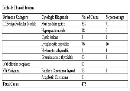
Results: Thyroid lesions formed 61.5 % (475) of the total number of head and neck masses. 99% of the cases were benign lesions. Lymphadenopathies formed 34 % (262) of the total number of head and neck masses. Most common diagnoses were reactive lymphadenitis (43.5%) followed by granulomatous lymphadenitis (12.5%). Salivary gland lesions formed 4.5 % (35) of the total number of head and neck masses. Sialadenitis was the most common among the benign lesions (40 %) and pleomorphic adenoma was commonest among neoplasms (37 %).
Conclusion: FNAC is a reliable, safe, cost-effective and minimally invasive procedure. FNAC can be advised as a primary diagnostic tool.
Downloads
References
2. Shahid F, Mirza T, Mustafa S, Sabahat S, Sharafat S. An experiential status of fine needle aspiration cytology of head and neck lesions in a tertiary care scenario. Journal of Basic & Applied Sciences. 2010 Dec 1;6(2).
3. Orell SR, Sterrett GF, Whitaker D. Fine needle aspiration cytology. Churchill Livingstone; 2005 Jul 8.
4. Gray W, Kocjan G. Diagnostic Cytopathology: Expert Consult: Online and Print. Elsevier Health Sciences; 2010 May 24.
5. Bibbo M, Wilbur D. Comprehensive cytopathology. Elsevier Health Sciences; 2014 Sep 5.
6. Tariq N, Sadiq S, Kehar S, Shafiq M. Fine needle aspiration cytology of head and neck lesions-an experience at the JINNAH post graduate medical centre, Karachi. Pak J Otolaryngol. 2007;23:63-5.
7. Amatya BB, Joshi AR, Singh SK, Panth R, Basnet RB. A study of fine needle aspiration cytology of head and neck masses and their corroboration by histopathology. Post graduate medical journal of national academy of medical sciences. 2009;6(2).
8. Fatima S, Arshad S, Ahmed Z, Hasan SH. Spectrum of cytological findings in patients with neck lymphadenopathy-experience in a tertiary care hospital in Pakistan. Asian Pac J Cancer Prev. 2011 Jan 1;12(7):1873-5.
9. Mundasad B, Mcallister I, Carson J, Pyper PC. Accuracy of fine needle aspiration cytology in diagnosis of thyroid swellings. Internet J Endocrinol. 2006;2(2):23-5.
10. El-Hag IA, Chiedo LC, AlRayees FA, Kollur SM (2003): FNAC of Head and Neck masses, 7 years experience in a secondary care hospital . acta Cytol., 47 (3): 387-92. [PubMed]
11. Chauhan S, Rathod D, Joshi DS. FNAC of swellings of head and neck region. Indian Journal of applied basic medical sciences. 2011;13(17):1-6.

 OAI - Open Archives Initiative