
Histomorphological study of uterine leiomyomas and its variants with brief review of literature
Abstract
Background: Uterine mesenchymal tumours are a heterogeneous group of neoplasms that can frequently be diagnostically challenging. Most subtypes of leiomyoma are chiefly of interest in that they mimic malignancy in one or more respects.
Objective: To evaluate the histomorphological features of uterine leiomyomas and its variants.
Materials and methods: Total of 477 cases of uterine leiomyomas and its variants were analysed prospectively in a period of 2years during July 2010 to June 2012 to assess the various pattern of leiomyomas. Cases were studied in detail about complete history, clinical examination and other findings.
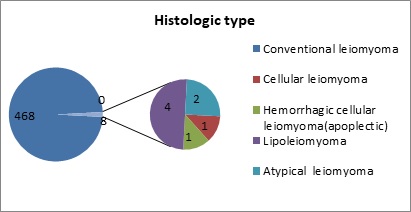
Results: In the study 468 (98.11%) cases showed features of conventional leiomyoma and 8 cases showed variants of leiomyomas (1.68%).
Conclusion: Although their diagnosis is straight forward in most cases, difficulties arise with particular leiomyoma variants, especially highly cellular leiomyoma (often confused with an endometrial stromal tumour) and leiomyoma with bizarre nuclei, mitotically active leiomyoma which may cause concern for leiomyosarcoma.
Downloads
References
2. Robboy SJ, McCluggage WG. Mesenchymal Uterine tumours, other than pure smooth muscle neoplasms, and adenomyosis. In: Robboy SJ, Mutter GM, Prat J, Bentley R, Russel P, Anderson MC editors. Textbook of Pathology of Female Reproductive Tract. 2ndedn, Elsevier, Churchill Livingstone 2009: 427-55.
3. Hendrickson MR, Tavassoli FA, Kempson RL, McCluggage WG, Haller U, Kubik- Hutch RA. Mesenchymal tumors and related lesions. In: Tavassoli FA, Deville P, editors. World Health Organization Classification of tumours. Pathology and genetics of Tumors of the Breast and Female Genital Organs. Lyon: IARC Press 2003; 233-44.
4. Robboy SJ, Bentley RC, Butnor K, Anderson MC. Pathology and pathophysiology of uterine smooth-muscle tumors. Environ Health Perspect. 2000; 108 (5):779-84.
5. Zaloudek CJ, Hendrickson MR, Saslow RA. Mesenchymal tumours of the Uterus. In: Kurman RJ, Elleson LH, Ronett BM. Blaustein`s Pathology of the Female Genital Tract. 6th ed, Springer 2011: 453-527.
6. Blom R, Guerrieri C, Stâl O, Malmström H, Simonsen E. Leiomyosarcoma of the uterus: A clinicopathologic, DNA flow cytometric, p53, and mdm-2 analysis of 49 cases. Gynecol Oncol. 1998; 68 (1): 54-61. doi:10.1006/ gyno.1997.4889
7. Gross KL, Morton CC. Genetics and the development of fibroids. Clin Obstet Gynecol. 2001;44(2):335-49.
8. Wantabe K, Suzukki T. Uterine leiomyoma versus leiomyosarcoma: a new attempt at differential diagnosis based on their cellular characteristics. Histopathol 2006; 48 (5):563-8.doi:10.1111/j.1365-2559.2006. 02368.x
9. Han SC, Kim MD, Jung DC, Lee M, Lee MS, Park SI, et al. Degeneration of leiomyoma in patients referred for uterine fibroid embolization: incidence, imaging features and clinical characteristics. Yonsei MedJ.2013;54(1):215-9.doi:10.3349/ymj.2013.54.1.215
10. Kurman RJ, Carcangiu ML, Herrington CS, YoungRH. WHO classification of tumours of female reproductiveorgans, 4th ed. Lyon, France: International Agency for Research on Cancer, 2014
11. Bell SW, Kempson RL, Hendrickson MR. Problematic uterine smooth muscle neoplasms. A clinicopathologic study of 213 cases. Am J Surg Pathol. 1994;18(6):535-58.
12. Boyd C, McCluggage WG. Unusual morphological features of uterine leiomyomas treated with progestogens. J Clin Pathol. 2011;64(6):485-9. doi: 10. 1136/jcp. 2011.089664. Epub 2011 Mar 11.
13. Croce S, Young RH, Oliva E. Uterine leiomyomas with bizarre nuclei: a clinicopathologic study of 59 cases. Am J Surg Pathol. 2014;38(10):1330-9. doi: 10.1097/PAS.0000000000000249.
14. Ip PP, Cheung AN. Pathology of uterine leiomyosarcomas and smooth muscle tumours of uncertain malignant potential. Best Pract Res Clin Obstet Gynaecol 2011; 25(6):691-704. doi: 10.1016/j. bpobgyn. 2011.07.003
15. Oliva E, Young RH, Clement PB, Bhan AK, Scully RE. Cellular benign mesenchymal tumors of the uterus. A comparative morphologic and immunohistochemical analysis of 33 highly cellular leiomyomas and six endometrial stromal nodules, two frequently confused tumors. Am J Surg Pathol. 1995;19(7):757-68.
16. Anderson MC. Female genital tract. In: Symmer SW ed. Systemic Pathology, 3rd ed, Edinburg, Churchill Livingstone, 1991.
17. Hendrickson MR and Kempson RL. Pure mesen-chymal tumours of the uterine corpus. In: Fox H Editors Obstetrical & Gynecological Pathology. 4th Ed. New York; Churchill Livingstone 1995: 511-86.
18. Jaiswal CJ. Vaginal management of uterocervical-myomas. J Obstet GynecolIndia 1996; 46: 260-3. DOI: http:// dx.doi.org/10.18203/ 2320-1770.ijrcog 20173500
19. Pujani M, Jairajpuri ZS, Rana S, Jetley S, Hassan MJ, Jain R. Cellular leiomyoma versus endometrial stromal tumor:A pathologists' dilemma. J Midlife Health.2015;6(1):31-4.doi:10.4103/0976-7800.153619

 OAI - Open Archives Initiative