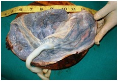
Morphological study of placenta in hypertensive disorders in pregnancy
Abstract
Introduction: Hypertensive disorders are common complications of pregnancy. Thorough macroscopic and microscopic examination of the placenta provides much insight into the prenatal health of the baby and the mother.
Objectives: 1. To study the morphological changes in the placenta in pregnant mothers. 2. Comparative study of morphological changes in the placenta among hypertensive and normotensive pregnant mothers.
Methods: An Observational Prospective Cohort Study was performed. Detail clinical history taken and placentae were collected from both 40 hypertensive and 40 normotensive mother's delivered in labour room or operation theatre. Both macroscopical and histopathological examination was done. Findings were recorded and analyzed statistically.
Results: The comparison of placental diameter, placental thickness, mean placental weight, placental volume, placental surface area between hypertensive and normotensive group showed statistically significant difference (p value < 0.05). Incidence of placental haematoma, infarction, basement membrane thickening of villi and syncytial knot in hypertensive group was 20%, 27.5%, 50% and 92.5% & in normotensive group was 5%, 10%, 12 % and 60% respectively. All cases in hypertensive group had placental fibrinoid necrosis of villi in comparison to 57.5% cases in normotensive group (p < 0.05). For fibrosis of villi and cytotrophoblatic proliferation p value was < 0.05 which was statistically significant.
Conclusion: Effects of hypertensive disorder in pregnancy reflect in gross and microscopic findings of placenta which may contribute to the further management of mother and baby.
Downloads
References
2. Barton JR, O'brien JM, et al. Mild gestational hypertension remote from term: progression and outcome. Am J Obstet Gynecol. 2001 Apr;184(5):979-83. DOI:10.1067/mob.2001.112905.[pubmed]
3. Salgado SS, Pathmeswaran A. Effects of placental infarctions on the fetal outcome in pregnancies complicated by hypertension. J Coll Physicians Surg Pak. 2008 Apr;18(4):213-6. doi: 04.2008/JCPSP.213216.[pubmed]
4. Udainia A, Jain ML. Morphological study of placenta in pregnancy induced hypertension with its clinical relevance. Journal of the Anatomical Society of India. 2001 Jun; 50(1): 24-7.
5. Udainia A. Bhagwat SS, Mehta CD. Relation between placental surface area, infarction and foetal distress in pregnancy induced hypertension with its clinical relevance. J Anat. Soc. Ind 2004; 53(1): 27-30.
6. Browne JC, Veall N. The maternal placental blood flow in normotensive and hypertensive women. J Obstet Gynaecol Br Emp. 1953 Apr;60(2):141-7.[pubmed]
7. Landesman R, Douglas RG, Holze E. The bulbar conjunctival vascular bed in the toxemias of pregnancy. Am J Obstet Gynecol. 1954 Jul;68(1):170-83. DOI:10.1016/0002-9378(54)90476-7.[pubmed]
8. Thomson AM, Billewicz WZ, Hytten FE. The weight of the placenta in relation to birthweight. J Obstet Gynaecol Br Commonw. 1969 Oct;76(10):865-72.[pubmed]
9. Stock MK, Anderson DF, Phernetton TM, et al. Vascular response of the fetal placenta to local occlusion of the maternal placental vasculature. J Dev Physiol. 1980 Oct;2(5):339-46.
10. Majumdar S, Dusguspta H, Bhattacharya K, Bhattcharya A. A study of placenta in normal & hypertensive pregnancies. J Anat. Soc. India 2005; 54(2): 1-9.
11. Wentworth P. Placental infarction and toxemia of pregnancy. Am J Obstet Gynecol. 1967 Oct 1;99(3):318-26. DOI:10.1016/s0002-9378(16)34537-9.[pubmed]
12. Joseph F Yetter III. Col, MC, USA, ‘American family Physician’ march 1998; Vol. 57 / No. 5 Published by American Academy Of Family Physician.
13. Benirschk. American Journal of Obstetrics and Gynaecology .1,New York 961;84:1595.
14. Shah R K et al. Journal of Obstetrics and Gynecology of India 1985;35:1809.
15. Shanklin D R. Journal of Obstetrics and Gynecology of the British Commonwealth 1958; 11: 129.
16. Patricia C. Gray’s Anatomy. 38th ed. New York: Churchill Livingstone,
17. Damania KR, Salvi VS, Ratnaparki SK, Daftari SN. The placenta in hypertensive disorder in pregnancy. J Obst and Gynaecol Ind 1989; 39: 28-31.
18. Fox H; The placenta in intra uterine growth retardation. In Ward RHT, Smith SK, Donnai D (Eds). Early foetal growth and development. RCOG Press, London: 1994. p. 223-235.
19. Kalousek DK, Langlosis S. The effects of placental and somatic chromosomal mosaicism on foetal growth. In. Ward RHT, Smith SK, Donnai (eds), Early foetal growth and development, RCOG Press; 1994. p. 245-256.
20. Mardi K, Sharma J. Histopathological evaluation of placentas in IUGR pregnancies. Indian J Pathol Microbiol. 2003 Oct;46(4):551-4.[pubmed]
21. Salgado SS, Angunawela P, Sirisena J, De Tissera A. Villous syncytial knots in hypertensive placentae. Sri Lanka J Obstet Gynaecol 2004; 26:33-7.
22. Fox H. Pathology of the Placenta. 2nd ed. W B: Saunders, London; 1997a
23. Benirschke K & Kaufmann P. ‘Pathology of Human Placenta’ . 2nd ed. Newyork: Springer Verlag;1990. P. 130.
24. Soma H, Yoshida K, Mukaida T, et al. Morphologic changes in the hypertensive placenta. Contrib Gynecol Obstet. 1982;9:58-75.[pubmed]
25. Jones CJP, Fox H. An ultrastructural and ultrahistochemical study of the human placenta in maternal pre-eclampsia. Placenta 1980; 1:61-76.[pubmed]
26. Damjanov I, Linder J. Andersons Pathology.10th ed. New York: Mosby; 1906. p. 2321.
27. Tinney B Parker F. The placenta in Toxaemia of pregnancy. AmJ Obstet Gynecol 1940; 39: 1000-1005.


 OAI - Open Archives Initiative


