
Cytomorphological patterns of cervical Papanicolaou smear abnormalities based on 2014 Bethesda System in North Karnataka region
Abstract
Introduction: Cervical PAP smears are a cost effective, out-patient procedure to screen patients for cervical pathology.
Objectives: To utilize cervical PAP smear examination in categorizing lesions according to the 2014 Bethesda System for cervical cytology, to analyse the spectrum of lesions andto evaluate its effectiveness as a screening procedure for detection of epithelial abnormalities in a teaching hospital in North Karnataka.
Methods: A prospective one year study was carried on all conventional PAP smears received in the Department of Pathology, Navodaya Medical College, Raichur. Reporting was done in accordance with the 2014 Bethesda System for reporting cervical cytology. Correlation was done with the clinical findings.
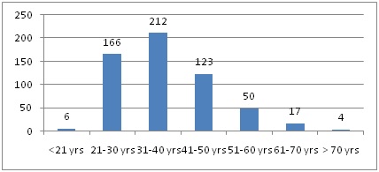
Results: A total of 578 cases were included in the study. The most common presenting complaint was abdominal pain (28.5%). The most frequent examination finding was white discharge per vaginum, WDPV (38.9%). 90.8% of PAP smears were categorized as ‘Negative for intraepithelial Lesion or Malignancy’. Specific infections were seen in 8.1%, squamous metaplasia in 15.5% and atrophic changes in 3.2%. Epithelial cell abnormalities comprised 9.2% of cases, of which Atypical Squamous Cells of Undetermined Significance (ASC-US) was 3.2%, Low grade Squamous Intraepithelial Lesion (LSIL) was 3.6%, High grade Squamous Intraepithelial Lesion (HSIL) was 1.9%, Squamous Cell Carcinoma (SCC) was 0.2% and Atypical Glandular Cells– Not Otherwise Specified (AGC-NOS) was 0.9%.
Conclusion: The overall prevalence of epithelial cell abnormalities concurred with studies done in other parts of India and constituted9.2% of the total smears screened, LSIL being the most common lesion.
Downloads
References
2. Bruni L, Albero G, Serrano B, Mena M, Gomez D, Munoz J, et al. ICO/IARC Information Centre on HPVand Cancer (HPV Information Centre). Human Papillomavirus and Related Diseases in India. Summary Report. 2018.
3. Sreedevi A, Javed R, Dinesh A. et al. Epidemiology of cervical cancer with special focus on India. Int J Womens Health. 2015 Apr 16;7:405-14. doi: 10.2147/IJWH.S50001. eCollection 2015.[pubmed]
4. Verma I, Jain V, Kaur T. Application of bethesda system for cervical cytology in unhealthy cervix. J Clin Diagn Res. 2014 Sep;8(9):OC26-30. doi: 10.7860/JCDR/2014/9620.4893. Epub 2014 Sep 20.[pubmed]
5. Bal MS, Goyal R, Suri AK, et al. Detection of abnormal cervical cytology in Papanicolaou smears. J Cytol. 2012 Jan;29(1):45-7. doi: 10.4103/0970-9371.93222.[pubmed]
6. Chichareon SB. Management of pre-invasive cervical cancer in low-resource setting. J Med Assoc Thai. 2004 Oct;87 Suppl 3:S214-22.[pubmed]
7. Kumari A, Pankaj S, Choudhary V, et al. Retrospective analysis of patients of cervical cancer a tertiary center in Bihar. Indian J Cancer. 2018 Jan-Mar;55(1):70-73. doi: 10.4103/ijc.IJC_482_17.[pubmed]
8. Foley G, Alston R, Geraci M, et al. Increasing rates of cervical cancer in young women in England: an analysis of national data 1982-2006. Br J Cancer. 2011 Jun 28;105(1):177-84. doi: 10.1038/bjc.2011.196. Epub 2011 Jun 7.[pubmed]
9. Alakananada, Sarma U, Biswas I. Histopathological Correlation with Cervical Cytology. IOSR J Dent Med Sci [Internet]. 2016;15(11):53–8. Available from: www.iosrjournals.org
10. Nayar R, Wilbur DC, editors. The Bethesda System for Reporting Cervical Cytology. Third. Springer International Publishing; 2015. 321 p.
11. Nayar R, Wilbur DC. The Pap Test and Bethesda 2014. "The reports of my demise have been greatly exaggerated." (after a quotation from Mark Twain). Acta Cytol. 2015;59(2):121-32. doi: 10.1159/000381842. Epub 2015 May 19.[pubmed]
12. Narayana G, Suchitra MJ, Sunanda G, et al. Knowledge, attitude, and practice toward cervical cancer among women attending Obstetrics and Gynecology Department: A cross-sectional, hospital-based survey in South India. Indian J Cancer. 2017 Apr-Jun;54(2):481-487. doi: 10.4103/ijc.IJC_251_17.[pubmed]
13. Sachan PL, Singh M, Patel ML, et al. A Study on Cervical Cancer Screening Using Pap Smear Test and Clinical Correlation. Asia Pac J Oncol Nurs. 2018 Jul-Sep;5(3):337-341. doi: 10.4103/apjon.apjon_15_18.[pubmed]
14. Verma A, Verma S, Vashist S, Attri S, Singhal A. A study on cervical cancer screening in symptomatic women using Pap smear in a tertiary care hospital in rural area of Himachal Pradesh, India. Middle East Fertil Soc J [Internet]. 2017;22(1):39–42. Available from: http://dx.doi.org/10.1016/j.mefs.2016.09.002
15. Saslow D, Solomon D, Lawson HW, et al. American Cancer Society, American Society for Colposcopy and Cervical Pathology, and American Society for Clinical Pathology screening guidelines for the prevention and early detection of cervical cancer. J Low Genit Tract Dis. 2012 Jul;16(3):175-204. DOI:10.1097/LGT.0b013e31824ca9d5.[pubmed]
16. Kulkarni PR, Rani H, Vimalambike MG, et al. Opportunistic screening for cervical cancer in a tertiary hospital in Karnataka, India. Asian Pac J Cancer Prev. 2013;14(9):5101-5.[pubmed]
17. Vaghela BK, Vaghela VK, Santwani PM, Vaghela BK. Analysis of abnormal cervical cytology in Papanicolaou smears at tertiary care center – A retrospective study. Int J Biomed Adv Res. 2014;5(1):47–9.
18. Nandwani RR, Totade S, Krishnan MG. Cytomorphological evaluation of squamous cell abnormalities observed on cervical smears in government medical college , Jabalpur , India : a five year study. Int J Res Med Sci. 2016;4(3):794–9.
19. Elhakeem HA, Al-Ghamdi AS, Al-Maghrabi JA. Cytopathological pattern of cervical Pap smear according to the Bethesda system in Southwestern Saudi Arabia. Saudi Med J. 2005 Apr;26(4):588-92.[pubmed]
20. Altaf FJ, Mufti ST. Pattern of cervical smear abnormalities using the revised Bethesda system in a tertiary care hospital in Western Saudi Arabia. Saudi Med J. 2012 Jun;33(6):634-9.[pubmed]
21. Maleki A, Ahmadnia E, Avazeh A, et al. Prevalence of Abnormal Papanicolaou Test Results and Related Factors among Women Living in Zanjan, Iran. Asian Pac J Cancer Prev. 2015;16(16):6935-9.[pubmed]
22. Bukhari MH, Saba K, Qamar S, et al. Clinicopathological importance of Papanicolaou smears for the diagnosis of premalignant and malignant lesions of the cervix. J Cytol. 2012 Jan;29(1):20-5. doi: 10.4103/0970-9371.93213.[pubmed]

 OAI - Open Archives Initiative