
Histopathological study of Non-neoplastic & Neoplastic ovarian lesions in a tertiary care hospital in Gujarat, India
Abstract
Aims & Objectives:(1) To know about various histopathological types of ovarian lesions presented and diagnosed at our institute. (2) To study the incidence of ovarian lesions with respect to patient’s age. (3) To study the frequency of ovarian lesions in terms of non-neoplastic or neoplastic, benign or malignant, unilateral or bilateral, etc.
Materials & Methods: The present study was performed at the Department of Pathology, GMERS Medical College & Hospital-Junagadh (Gujarat, India) from January 2015 to December 2018 and includes 100 cases of ovarian lesions diagnosed on both clinical & histopathological basis. We have received ovarian specimens and performed routine grossing and H& E staining procedure. We have included parameters like Age wise incidence, Nature of Lesion, Frequency & Laterality in this present study.
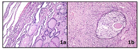
Results: Out of100 cases, 89% are unilateral and 11% are bilateral. 52% lesions are Benign Neoplasms, 44% lesions are Non-neoplastic Cysts and 4% lesions are Borderline & Malignant Neoplasms. Majority of cases (58%) belong to age group of 20-39 years. Among Non-neoplastic Lesions, Follicular Cyst is common & frequently bilateral while among Benign Neoplasms, Serous Cystadenoma is common & frequently bilateral.
Conclusion: Ovarian Lesions both non-neoplastic and neoplastic include a variety of morphological features and show a particular age wise incidence. Role of histopathological evaluation remains always important in both diagnosis & management of such cases, particularly in cases of Malignant Lesions in order to save the patient’s life.
Downloads
References
2. Modi D, Rathod GB, Delwadia KN, Goswami HM. Histopathological pattern of neoplastic ovarian lesions. IAIM. 2016; 3(1):51-7.
3. Vinay Kumar, Abul K. Abbas, Jon C. Aster.pathology of female genital tract, Robbins and Cotran Pathological Basis of Disease,9thedition(II). Elsevier;2015(22):1023.
4. Prakash A, Chinthakindi S, Duraiswami R, Indira V. Histopathological study of ovarian lesions in a tertiary care center in Hyderabad, India: a retrospective five-year study Int J Adv Med. 2017; 4(3):745-49.
5. Gurung P, Hirachand S, Pradhanang S. Histopathological study of ovarian cystic lesions in Tertiary Care Hospital of Kathmandu, Nepal. Journal of Institute of Medicine 2013, 35(3):44-47.
6. Prabhakar BR, Maingi K. Ovarian tumours--prevalence in Punjab. Indian J Pathol Microbiol. 1989 Oct;32(4):276-81.[pubmed]
7. Couto F, Nadkarni NS, Rebello MJ. Ovarian tumors in Goa. A Clinicopathological study. J ObstetGynecol of India. 1993; 43(3):408-12.
8. Zaman S, Majid S, Hussain M, et al. A retrospective study of ovarian tumours and tumour-like lesions. J Ayub Med Coll Abbottabad. 2010 Jan-Mar;22(1):104-8.[pubmed]
9. Yogambal M, Arunalatha P, Chandramouleeswari K, Palaniappan V. Ovarian tumors - Incidence and distribution in a tertiary referral center in South India. IOSR J Dent Med Sci. 2014; 13(2):1400-3.
10. Pudasaini S, Lakhey M, Hirachand S, et al. A study of ovarian cyst in a tertiary hospital of Kathmandu valley. Nepal Med Coll J. 2011 Mar;13(1):39-41.[pubmed]
11. Maliheh A, et al. Surgical Histopathology of Benign Ovarian Cysts: A Multicenter Study. Iranian Journal of Pathology 2010; 5(3): 132-6.
12. Mondal SK, Et al. Histologic pattern, bilaterality and clinical evaluation of 957 ovarian neoplasms: a 10 years study in a tertiary hospital of Eastern India. J Can Res Ther2011;7:433-7.
13. Iqbal J et al. Pattern of Ovarian Pathologies. Journal of Rawalpindi Medical College (JRMC) 2013;17(1): 113-5.
14. Yasmin S, Yasmin A, Asif M. Clinicohistological pattern of ovarian tumours in Peshawar region. J Ayub Med Coll Abbottabad. 2008 Oct-Dec;20(4):11-3.[pubmed]
15. Ramachandran G, Harilal KR, Chinnamma K, Thangavelu H. Ovarian neoplasms -A study of 903 cases. J ObstetGynecol India 1972; 22:309 -15.
16. Pilli GS, Suneeta KP, Dhaded AV, et al. Ovarian tumours: a study of 282 cases. J Indian Med Assoc. 2002 Jul;100(7):420, 423-4, 447.[pubmed]
17. Kar T, Kar A, Mohapatra PC. Intra-operative cytology of ovarian tumors. J ObstetGynecol India. 2005; 55(4):345-9.
18. Hatwal D, Choudhari S, Batra N, Bhatt P, Bhatt S. Clinico-histopathological analysis of neoplastic and non-neoplastic lesion of ovary in Garhwal region of Uttarakhand: A 4 year study at tertiary level hospital. Indian Journal of Pathology and Oncology, April-June 2016; 3(2):133-140.

 OAI - Open Archives Initiative