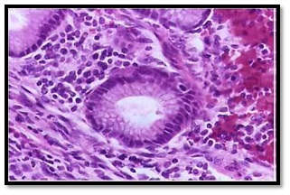
Retrospective Study of histomorphological spectrum of ovarian tumours in GCS Medical College, Hospital and Research Center, Ahmedabad
Abstract
Background and objectives: Ovarian tumours accounts for 30% of female genital cancers and have become increasingly important not only because of the large variety of neoplastic entities but also because they have gradually increased the mortality rate. Ovarian neoplasm remains asymptomatic until there is massive ovarian enlargement which causes compression of pelvic structures, ascites, abdominal distension or distant metastasis.Thus present study was undertaken to analyze the frequency of various histomorphological spectrum, histological subtypes and age distribution pattern of ovarian tumours.
Methods: Retrospective study was carried during period of 1st June 2017 to 30st November 2018, 57 cases of ovarian neoplastic lesions were taken from the records of the department.
Results: A total number of 57 cases were studied. Among these, 41cases (71.92%) were of benign tumours, 2 cases (3.50%) were of borderline tumours and 14 cases (24.56%) were of malignant tumours. Serous cystadenomas (26/41) (63.41%) were the commonest benign tumours followed by Mucinous cystadenomas (6/41)(14.63%). Among the malignant surface epithelial tumours serous cystadenocarcinomas (6/14) (42.86%) were most common followed by Mucinous cystadenocarcinomas (3/14) (21.43%). Germ cell neoplasms constituted 19.30% of all ovarian neoplasms.
Conclusion: Ovarian tumours cannot easily be diagnosed by clinical symptoms only, histological examination is therefore necessary for diagnosis and grading of ovarian tumours. Females with family history of endometrial and breast carcinoma has got increased incidence of ovarian carcinoma and ovary is also common site for metastasis from other primary gynecological as well as non gynecological carcinomas e.g. endometrium, stomach and breast. Hence present study was carried out in the department of pathology for early diagnosis of ovarian carcinomas, their histological sub typing and their age distribution pattern.
Downloads
References
2. Shirish S Chandanwale, Rahul Jadhav, Ruby Rao, PiyushaNaragude, Sunita Bhamnikar, Jehan Nizam Ansari. Clinicopathologic Study of Malignant Ovarian Tumors: A Study of Fifty Cases. Med J DY Patil Univ 2017;10:430-7.
3. Bhattacharya M, Shinde SD, Purandare VN. A clinicopathological analysis of 270 ovarian tumours. J Postgrad Med. 1980 Apr;26(2):103-7.
4Susan J. Ramus and Simon A. Gayther .The Contribution of BRCA1 and BRCA2 to Ovarian CancerMol Oncol. 2009 Apr; 3(2): 138–150.
5. Schmeler KM, Lynch HT, Chen LM, et al. Prophylactic surgery to reduce the risk of gynecologic cancers in the Lynch syndrome. N Engl J Med. 2006 Jan 19;354(3):261-9.[pubmed]
6. Isabel Alvarado-Cabrero, Adriana Rodríguez-Gómez, Jorge Castelan-Pedraza, and Raquel Valencia-Cedillo, Metastatic Ovarian Tumors A Clinicopathologic Study of 150 Cases.Analytical and quantitative cytology and histopathology35(5):241-248 October 2013.
7. Lee SJ, Bae JH, Lee AW, et al. Clinical characteristics of metastatic tumors to the ovaries. J Korean Med Sci. 2009 Feb;24(1):114-9. doi: 10.3346/jkms.2009.24.1.114. Epub 2009 Feb 28.[pubmed]
8. Shirish S Chandanwale, Rahul Jadhav, Ruby Rao, PiyushaNaragude, Sunita Bhamnikar, Jehan Nizam Ansari.Clinicopathologic Study of Malignant Ovarian Tumors: A Study of Fifty Cases.Oncology (Williston Park). 2004 May;18(5):645-53, discussion 653-4, 656, 658.
9. Laury AR, Hornick JL, Perets R, et al. PAX8 reliably distinguishes ovarian serous tumors from malignant mesothelioma. Am J Surg Pathol. 2010 May;34(5):627-35. doi: 10.1097/PAS.0b013e3181da7687.[pubmed]
10. Digant Gupta and Christopher G Lis.Role of CA125 in predicting ovarian cancer survival - a review of the epidemiological literature J Ovarian Res. 2009; 2: 13.[pubmed]
11. Russell Vang, Ie-Ming Shih, and Robert J. Kurman, Ovarian low-grade and high-grade serous carcinoma: Pathogenesis, Clinicopathologic and Molecular Biologic Features, and Diagnostic Problems Published in final edited form as: Adv AnatPathol. 2009 Sep; 16(5): 267–282.
12. Chiesa AG, Deavers MT, Veras E, et al. Ovarian intestinal type mucinous borderline tumors: are we ready for a nomenclature changeInt J GynecolPathol. 2010 Mar;29(2):108-12.[pubmed]
13. Cathro HP, Stoler MH. Expression of cytokeratins 7 and 20 in ovarian neoplasia. Am J Clin Pathol. 2002 Jun;117(6):944-51. DOI:10.1309/2T1Y-7BB7-DAPE-PQ6L.[pubmed]
14. Hackethal A, Brueggmann D, Bohlmann MK, et al. Squamous-cell carcinoma in mature cystic teratoma of the ovary: systematic review and analysis of published data. Lancet Oncol. 2008 Dec;9(12):1173-80. doi: 10.1016/S1470-2045(08)70306-1.[pubmed]
15. S. F. Serov R. E. Scully .Histological typing of ovarian tumours :international histological classification of tumours no.9 world health organization, geneva, switzerland
16. Pilli GS, Suneeta KP, Dhaded AV, et al. Ovarian tumours: a study of 282 cases. J Indian Med Assoc. 2002 Jul;100(7):420, 423-4, 447.[pubmed]
17. Guppy AE, Nathan PD, Rustin GJ. et al. Epithelial ovarian cancer: a review of current management. Clin Oncol (R Coll Radiol). 2005 Sep;17(6):399-411.[pubmed]
18. Yasmin S, Yasmin A, Asif M. Clinicohistological pattern of ovarian tumours in Peshawar region. J Ayub Med Coll Abbottabad. 2008 Oct-Dec;20(4):11-3.[pubmed]
19. Tyagi SP, Maheswari V, Tyagi N, et al. Solid tumours of the ovary. J Indian Med Assoc. 1993 Sep;91(9):227-30.[pubmed]
20. Ahmad Z, Kayani N, Hasan SH, et al. Histological pattern of ovarian neoplasma. J Pak Med Assoc. 2000 Dec;50(12):416-9.[pubmed]


 OAI - Open Archives Initiative


