
Histopathological study of meningioma in a tertiary care centre: a two years experience
Abstract
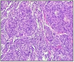
Background: Meningioma is a neoplasm arising from the arachnoidal cap cells in the meningeal coverings of the spinal cord and brain. These are the most common benign intracranial tumours and account for about 13-26% of all primary brain neoplasms. These are generally benign neoplasms, but about 10% are atypical or malignant.
Objective: To study the variants and histopathological spectrum of meningioma. Material & Methods: This study includes 29 cases of meningioma diagnosed over a period of two years.
Result: Most common variant was noted to be meningothelial meningioma followed by transitional meningioma. Out of the 29 cases, grade I were 96.55% whereas grade II were 3.44%.
Conclusion: From our study, we conclude that meningothelial meningioma is the most common variant. Benign meningiomas are the most common type.
Downloads
References
2. Louis DN, Ohgaki H, Wiestler OD, Cavenee WK. WHO Classification of Tumours of the Central Nervous System. IARC: Lyon. IARC: Lyon 2007. 2007. 164-172 p.[pubmed]
3. Modha A, Gutin PH. Diagnosis and treatment of atypical and anaplastic meningiomas: A review. Vol. 57, Neurosurgery. 2005. p. 538–49.[pubmed]
4. Backer-Grøndahl T, Moen BH, Torp SH. The histopathological spectrum of human meningiomas. Int J Clin Exp Pathol. 2012;5(3):231–42.[pubmed]
5. Prayson RA. Pathology of Meningiomas. In: Meningiomas [Internet]. London: Springer London; 2009 [cited 2016 Dec 23]. p. 31–43. Available from: http://link.springer.com/10.1007/978-1-84628-784-8_5
6. Basu K, Majumdar K, Chatterjee U, Ganguli M, Chatterjee S. En plaque meningioma with angioinvasion. Indian J Pathol Microbiol [Internet]. 2010;53(2):319–21. Available from: http://www.ncbi.nlm.nih.gov/entrez/query.fcgi?cmd=Retrieve&db=PubMed&dopt=Citation&list_uids=20551544.[pubmed]
7. Paik SS, Jang SJ, Park YW, Hong EK, Park MH, Lee JD. Microcystic meningioma - A case report. Vol. 11, Journal of Korean Medical Science. 1996. p. 540–3.[pubmed]
8. Mahmood A, Caccamo D V, Tomecek FJ, Malik GM. Atypical and malignant meningiomas: a clinicopathological review. Neurosurgery [Internet]. 1993;33(6):955–63. Available from: http://meta.wkhealth.com/pt/pt-core/template-journal/lwwgateway/media/landingpage.htm?issn=0148-396X&volume=33&issue=6&spage=955
9. Louis DN, Perry A, Reifenberger G, von Deimling A, Figarella-Branger D, Cavenee WK, et al. The 2016 World Health Organization Classification of Tumors of the Central Nervous System: a summary. Vol. 131, Acta Neuropathologica. 2016. p. 803–20.[pubmed]
10. Shah S, Gonsai RN, Makwana R. HISTOPATHOLOGICAL STUDY OF MENINGIOMA IN CIVIL HOSPITAL , AHMEDABAD. Int J Curr Res Rev. 2013;5(3):76–82.
11. Gadgil NM, Margam SR, Chaudhari CS, Kumavat PV. The histopathological spectrum of meningeal neoplasms. Indian J Pathol Oncol [Internet]. 2016;3(3):432–6. Available from: http://www.indianjournals.com/ijor.aspx?target=ijor:ijpo&volume=3&issue=3&article=014
12. Reddy R, Singh R. Histopathological spectrum of meningioma and its variants. Asian Pacific J Heal Sci. 2016;3(1):151–5.
13. Lakshmi SS. Meningiomas: A Clinicopathological study. Int J Med Res Heal Sci [Internet]. 2015;4(4):827–31. Available from: http://www.indianjournals.com/ijor.aspx?target=ijor:ijmrhs&volume=4&issue=4&article=021
14. Ruiz J, Martínez A, Hernández S, Zimman H, Ferrer M, Fernández C, et al. Clinicopathological variables, immunophenotype, chromosome 1p36 loss and tumour recurrence of 247 meningiomas grade I and II. Histol Histopathol. 2010;25(3):341–9.
15. Desai P, Patel D. Histopathological study of meningioma. Int J Med Sci Public Heal [Internet]. 2016;5(2):327–30. Available from: http://www.scopemed.org/?mno=206109
16. Bansal D, Diwaker P, Gogoi P, Nazir W, Tandon A. Intraparenchymal angiomatous meningioma: A diagnostic dilemma. J Clin Diagnostic Res. 2015;9(10):ED07-ED08.
17. Wang DJ, Xie Q, Gong Y, Wang Y, Cheng HX, Mao Y, et al. Secretory meningiomas: Clinical, radiological and pathological findings in 70 consecutive cases at one institution. Int J Clin Exp Pathol. 2013;6(3):358–74.
18. Regelsberger J, Hagel C, Emami P, Ries T, Heese O, Westphal M. Secretory meningiomas: a benign subgroup causing life-threatening complications. Neuro Oncol. 2009;11:819–24.
19. Louis DN, Ohgaki H, Wiestler OD, Cavenee WK, Burger PC, Jouvet A, et al. The 2007 WHO classification of tumours of the central nervous system. Vol. 114, Acta Neuropathologica. 2007. p. 97–109.[pubmed]
20. Riemenschneider MJ, Perry A, Reifenberger G. Histological classification and molecular genetics of meningiomas. Lancet Neurol. 2006;5(12):1045–54.[pubmed]
21. Taghipour M, Rakei SM, Monabati A. The role of estrogen and progesterone receptors in grading of the malignancy of meningioma. Iran Red Crescent Med J. 2007;9(1):17–21.

 OAI - Open Archives Initiative