
Histomorphological spectrum of eyelid lesions–A 6 year retrospective study
Abstract
Background: Eyelid lesions are encountered by all primary care physicians and Ophthalmologists. Histology of eyelid comprises various components and structures that give rise to a wide spectrum of pathologies. The clinical presentation of eyelid lesions is myriad with benign lesions masquerading malignant tumours. Though eyelid lesions are fairly common in Indian subcontinent, there is paucity of reports in Indian literature. This study was undertaken to characterize the distribution of various eyelid lesions and clinicopathological correlation in a tertiary care centre of South India.
Objectives: To retrospectively carry out a clinicopathological analysis of eyelid lesions requiring surgical excision in the Department of Pathology of a tertiary care centre in South India.
Methods: A retrospective review of clinicopathological profile of excised eyelid lesions diagnosed in our tertiary care centre was done. Clinicopathological data were retrieved from patient’s clinical records and biopsy reports.
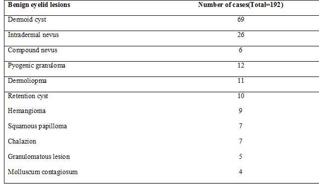
Result: Among 219 eyelid lesions, 192 were benign lesions and 27 were malignant tumours. The most common eyelid lesion was dermoid cyst (69 cases) followed by Nevus (32 cases) and Pyogenic Granuloma (12 cases). The most common malignant eyelid lesion was Sebaceous Cell Carcinoma (13 cases).
Conclusion: Dermoid cyst and Nevus are the most common eyelid lesions requiring biopsy and sebaceous cell carcinoma is the most common malignant eyelid tumour. All surgically excised eyelid lesions must be subjected to histopathological examination without fail to provide a definitive diagnosis, continued patient care and management.
Downloads
References
2. Mondal SK, Dutta TK. Cytohistological study of eyelid lesions and pitfalls in fine needle aspiration cytology. Journal of Cytology. 2008;25(4):133-7.
3. Sheikh IY, Shah FR, Gandhi MB, Shan CK, Shah NR, Ophthalmic neoplastic lesions. A retrospective study of 4 years. Gujarat Medical Journal.2012;67(2):53-7.
4. Kumar R, Adhikari R, Sharma M, Phokharel D, Gautam N. Patterns of ocular malignant tumors in Bhairahwa, Nepal. The Internet Journal of Ophthalmology and Visual science . 2008;7(1):1-6.
5. Sanjay CC, Shan SJ, Patel AB, Rathod HK, Surve SD, Nasit JG, A histopathological study ofophthalmic lesions at a teaching hospital. National J Medical Research. 2012;2(2):133-6.
6. Al-Faky YH. Epidemiology of benign eyelid lesions in patients presenting to a teaching hospital. Saudi J Ophthalmol. 2012 Apr;26(2):211-6. doi: 10.1016/j.sjopt.2011.05.005. Epub 2011 May 30.[pubmed]
7. Gupta P, Gupta RC, Khan L. Profile of eyelid malignancy in a Tertiary Health Care Center in North India. J Cancer Res Ther. 2017 Jul-Sep;13(3):484-486. doi: 10.4103/0973-1482.183215.[pubmed]
8. Mohan BP, Letha V. Profile of eye lid lesions over a decade: a histopathological study from a tertiary care center in South India. Int J Adv Med 2017;4:1406-11.
9. Abdi U, Tyagi N, Maheshwari V, et al. Tumours of eyelid: a clinicopathologic study. J Indian Med Assoc. 1996 Nov;94(11):405-9, 416, 418.[pubmed]
10. Tesluk GC. Eyelid lesions: incidence and comparison of benign and malignant lesions. Ann Ophthalmol. 1985 Nov;17(11):704-7.[pubmed]
11. Obata H, Aoki Y, Kubota S, et al. [Incidence of benign and malignant lesions of eyelid and conjunctival tumors]. Nippon Ganka Gakkai Zasshi. 2005 Sep;109(9):573-9.[pubmed]
12. Mihaela-Cristiana Coroi, Elena Rosca, Gabriela Mutiu, T.Coroi, Marinela Bonta. Eyelid tumors: histopathological and clinical study perfomed in County Hospital of Oradea between 2000-2007. Romanian Journal of Morphology and Embryology. 2010;51(1):111-5
13. Sean Paul, Dat TVo, RonaZ, Silkiss. Malignant and benign eyelid lesions in San Franscisco: Study of a diverse urban population. American Journal of Clinical Medicine. Winter2011;8(1):40-46
14. Ho M, Liu DT, Chong KK, et al. Eyelid tumours and pseudotumours in Hong Kong: a ten-year experience. Hong Kong Med J. 2013 Apr;19(2):150-5.[pubmed]
15. Ramya BS, Dayananda SB, Chinmayee JT, Raghupathi AR. Tumors of the eyelid- A Histopathological Study of 86 cases in a Tertiary Care Hospital. International Journal of Scientific and Research Publications,2014;4(11):1-5
16. Huang YY, Liang WH, Tsai CC, Kao SC, Yu WK, Kau HC, Liu JL. Comparison of the clinical characteristics and outcome of Benign and Malignant Eyelid Tumors: An analysis of 4521 Eyelid tumors in a Tertiary Medical Centre. Bio Med Research International.2015. doi : 10.1155/2015/453091
17. Anandini GM, Parikh SB, Shah NR. Histopathological Study of Eyelid Lesions. National Journal of Laboratory Medicine. 2018;7(1):07-11
18. Jahagirdar SS, Thakre TP, Kale SM, et al. A clinicopathological study of eyelid malignancies from central India. Indian J Ophthalmol. 2007 Mar-Apr;55(2):109-12.[pubmed]
19. Fouzia Farhat, Qamar Jamal, Mahmood Saeed, Zia Ghaffar. Evaluation of Eyelid Lesions at a Tertiary Care Hospital, Jinnah Postgraduate Medical Centre (JPMC), Karachi. Pak J Opthalmol 2010;26(2):83-6
20. Kale SM1, Patil SB, Khare N, et al. Clinicopathological analysis of eyelid malignancies - A review of 85 cases. Indian J Plast Surg. 2012 Jan;45(1):22-8. doi: 10.4103/0970-0358.96572.[pubmed]
21. Kafle, S., Khadka, D., Karki, S., & Lavaju, P. (2016). Spectrum of ocular malignant tumors in a tertiary care teaching hospital. Journal Of Patan Academy Of Health Sciences, 2016; 3(1), 15-17
22. Gupta Y, Gahine R, Hussain N, et al. Clinico-Pathological Spectrum of Ophthalmic Lesions: An Experience in Tertiary Care Hospital of Central India. J Clin Diagn Res. 2017 Jan;11(1):EC09-EC13. doi: 10.7860/JCDR/2017/23589.9230. Epub 2017 Jan 1.[pubmed]
23. Wang JK, Liao SL, Jou JR, et al. Malignant eyelid tumours in Taiwan. Eye (Lond). 2003 Mar;17(2):216-20.DOI:10.1038/sj.eye.6700231.[pubmed]
24. Kaliki S, Ayyar A, Dave TV, Ali MJ, Mishra DK, Naik MN (2015) Sebaceous gland carcinoma of the eyelid: clinicopathological features and outcome in Asian Indians. Eye (Lond) 2015; 29(7):958–963
25. Lin HY, Cheng CY, Hsu WM, et al. Incidence of eyelid cancers in Taiwan: a 21-year review. Ophthalmology. 2006 Nov;113(11):2101-7. Epub 2006 Sep 7.[pubmed]

 OAI - Open Archives Initiative