
Ovarian mass lesions: evaluation of ultrasound guided fine needle aspiration cytology with histopathological correlation
Abstract
Background: Ovarian masses are frequent finding in females of reproductive age group. Image-guided fine-needle aspiration cytology (FNAC) of ovarian lumps is being increasingly used for the successful diagnosis of ovarian tumors, although borderline cases may be difficult to diagnose by this method.
Objective: The present study was performed to evaluate the role of US-guided FNAC in pre-operative cytological diagnosis of ovarian masses in comparison with histopathology and to assess the pitfalls and limitations of cytological interpretation.
Materials and Methods: The study was conducted on 160 female patients. Diagnosis was established by FNAC performed under image guidance (ultrasonography/computed tomography) followed by histopathological examination. Cytologic diagnoses were compared with the histopathological diagnosis.
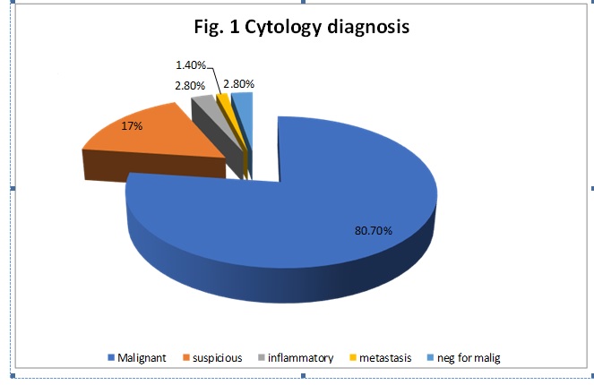
Results: On cytology and histopathology comparison, concordance was found to be 90.4% in case of malignancy, 94% in cases of suspicious for malignancy, 100% in cases of inflammatory lesions, 50 % in cases of metastasis. Chi-square test was performed and p value was statistically significant (p < 0.0001).
Conclusion: USG-guided FNAC seems to be a relatively safe, simple, fast and cost-effective procedure where most ovarian malignancies either present late in their course or no screening method is available. In addition this procedure may be useful in deciding management guidelines prior to any surgical intervention.
Downloads
References
2. Neetu Agarwal et al. Ovarian Neoplasm: Diagnostic Accuracy of Ultrasound Guided Fine Needle Aspiration Cytology with Histopathological Correlation. IOSR Journal of Dental and Medical Sciences,2014;13(7):24-28.
3. S Goel et al. Ultrasound Guided Fine Needle Aspiration Cytology in Ovarian Neoplasms: An Assessment of Diagnostic Accuracy and Efficacy and Role in Clinical Management. The Internet Journal of Pathology, 2010;11(2).
4. Scully RE. Tumours of the ovary and mal developed grounds. In Atlas of tumour pathology. 2nd series fascicle 16; Washington DC. Armed Forces Institute of Pathology 1981.
5. Anjali Bandyopadhyay et al. Fine needle aspiration cytology of ovarian tumors with histological correlation. Journal of Cytology, 2012;29(1).
6. Yang CY1, Kuo HW, Chiu HF. Age at first birth, parity, and risk of death from ovarian cancer in Taiwan: a country of low incidence of ovarian cancer. Int J Gynecol Cancer. 2007 Jan-Feb;17(1):32-6. DOI:10.1111/j.1525-1438.2007.00804.x.[pubmed]
7. Huusom LD, Frederiksen K, Høgdall EV, et al. Association of reproductive factors, oral contraceptive use and selected lifestyle factors with the risk of ovarian borderline tumors: a Danish case-control study. Cancer Causes Control. 2006 Aug;17(6):821-9. DOI:10.1007/s10552-006-0022-x.[pubmed]
8. Olsen CM, Cnossen J, Green AC, et al. Comparison of symptoms and presentation of women with benign, low malignant potential and invasive ovarian tumors. Eur J Gynaecol Oncol. 2007;28(5):376-80.[pubmed]
9. Ray et al. USG guided FNAC of ovarian mass lesions: A cyto-histopathological correlation, with emphasis on its role in pre-operative management guidelines. J Turk Ger Gynecol Assoc 2014;15:6-12.[pubmed]
10. Subrata Pal et al. Evaluation of Ultrasound-Guided Fine-Needle Aspiration Cytology of Ovarian Masses with Histopathological Correlation. Acta Cytologica 2015;59:149–155.
11. Robert V. Higgins et al. Comparison of fine-needle aspiration cytologic findings of ovarian cysts with ovarian histologic findings. Am J Obstet Gynecol 1999;180(3).
12. Ghazala Mehdi et al. Image-guided fine-needle aspiration cytology of ovarian tumors: An assessment of diagnostic efficacy. Journal of Cytology,2010;27(3).

 OAI - Open Archives Initiative