
Estimation of platelet counts: auto analyzer versus counts from peripheral blood smear based on traditional and platelet: red blood cell ratio method
Abstract
Introduction: Accurate platelet counts are of utmost importance in clinical practice The methods used for estimating platelet count are: Manual method using counting chamber, Examination of a peripheral blood smear (PBS) and automated hematology analyzers Automated hematology analyzers produce erroneous results in the presence of particles of similar size and/or light scatter like fragmented red blood cells (RBC), microcytic RBCs and in the presence of giant platelets and platelet clumps.
Aims and Objectives: Comparison of platelet count by three methods: 1. Automated six part analyzer. Traditional method: Counting average platelets per ten high power fields and multiplying the same by 15,000. 3. From smear by counting the number of platelets per 1000 RBCs and calculating the platelet count on the basis of platelet: RBC ratio.
Methods: Ethylene Diamine Tetra-acetic acid (EDTA) samples for platelet counts over a period of two months were analyzed by the above-mentioned methods. Statistical software SPSS 2 and independent T tests were used to compare the variables between the groups. Sensitivity and specificity of the methods was calculated.
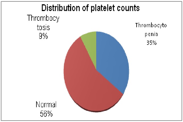
Results: 250 EDTA samples were analyzed of which normal platelet counts were (56.4%) thrombocytopenic (35.2%) and thrombocytosis (8.4%). There was no significant difference between the platelet counts done by the auto analyzer compared with traditional (P value 0.50) and platelet: RBC ratio method (0.906). Specificity of the methods was 99.1% and the sensitivity was 92.5%.
Conclusion: Platelet counts by traditional and platelet: RBC ratio can be used as alternate reliable methods as compared to the auto analyzers.
Downloads
References
2. Boulassel M-R, Al-Farsi R, Al-Hashmi S, Al-Riyami H, Khan H, Al-KindiS. Accuracy of platelet counting by optical and impedance methods in patients with thrombocytopenia and microcytosis. Sultan Qaboos Univ Med J. 2015;15(4):e463–8. doi: 10.18295/squmj.2015.15.04.004
3. Marionneaux S, Francisco N, Chan V, Hanenberg J, Rafael J, Chua C, et al. Comparison of automated platelet counts and potential effect on transfusion decisions in cancer patients. Am J Clin Pathol. 2013; 140 (5): 747–54.10. 1309/ AJCP58 INTITVGQZI
4. Brahimi M, Osmani S, Arabi A, et al. The estimation of platelet count from a blood smear on the basis of the red cell: platelet ratio. Turk J Heamtol2009;26 (1):21-4
5. Hutchinson RE, Mc Pherson RA, Schexneider KI. Basic examination of blood and bone marrow. In: Mc Pherson RA, Pincus MR, editors. Henry’s clinical diagnosis and management by laboratory methods.22nd ed. Philadelphia:Elsevier Saunders; 2011: 509-35.
6. Moreno A, Menke D. Assessment of platelet numbers and morphology in the peripheral blood smear. Clin Lab Med.2002;22(1):194-213
7. Rodak BF, Fritsma GA, Doig K. Hematology: Clinical principles and applications. 4th ed. Elsevier- Saunders; 2011; p. 202.
8. Malok M, Titchener EH, Bridgers C, Lee BY, Bamberg R Comparison of two platelet count estimation methodologies for peripheral blood smears. Clin Lab Sci. 2007 summer; 20 (3): 154-60.
9. UmaraniMK,Shashidhar HB. Estimation of platelet count from peripheral blood smear based on platelet: red blood cell ratio. A prospective study in a tertiary care hospital. Indian Journal of Pathology and Oncology, April-June 2016;3(2):351-353.10.5958/ 2394-6792. 2016.00066.1
10. Balduini, C. L.&Noris, P. Platelet count and aging. Haematologica 2014; 99: 953–955.
11. Al-Hosni ZS, Al-Khabori M, Al-Mamari S, et al. Reproducibility of Manual Platelet Estimation Following Automated Low Platelet Counts. Oman Medical Journal. 2016; 31(6):409-413. 10.5001/omj. 2016.83
12. Umashankar T, Thomas BM, Sahana P Estimation of platelet count in unstained peripheral blood smears in comparison with stained smears and evaluation of its efficacy. Estimation of platelet count in unstained peripheral blood smears in comparison with stained smears and evaluation of its efficacy. Malays J Pathol. 2014 Dec; 36(3):195-9.
13. Phakhounthong K, Chaovalit P, Jittamala P, BlacksellSD, Carter MJ, Turner P et al. Predicting the severity of dengue fever in children on admission based on clinical features and laboratory indicators: application of classification tree analysis. BMC Pediatrics. 2018; 18:109.doi:10. 1186/s 12887-018-1078-y.
14. MilovanovicAlempijevic T, StojkovicLalosevic M, Dumic I, et al. Diagnostic Accuracy of Platelet Count and Platelet Indices in Noninvasive Assessment of Fibrosis in Nonalcoholic Fatty Liver Disease Patients. Canadian Can J Gastroenterol Hepatol. 2017; 2017: 6070135. doi:10.1155/2017/ 6070135.
15. De la Salle BJ, McTaggart PN, Briggs C, Harrison P, Doré CJ, Longair I, et al. The accuracy of platelet counting in thrombocytopenic blood samples distributed by the UK National External Quality Assessment Scheme for General Haematology. Am J Clin Pathol. 2012 Jan;137 (1):65–74.doi.org/10. 1309/AJCP86JMBFUCFCXA
16. Buttarello M. Quality specification in hematology: the automated blood cell count. Clin Chim Acta. 2004;346:45–54. doi: 10.1016/j.cccn.2004.02.03
17. National Accreditation Board for Testing and Calibration Laboratories (IN). NABL 112 Specific Criteria for Accreditation of Medical Laboratories. Gurgaon: National Accreditation Board for Testing and Calibration Laboratories; 2016. p. 36
18. Jain A, Jain S, Singh N, et al. Storage stability of commonly used hematological parameters at 33 °C: Electronic supplementary material available online for this article. Biochem Med (Zagreb). 2018 Jun 15; 28(2): 020901
19. Bain BJ. Diagnosis from the blood smear. N Engl J Med. 2005;353:498-507 DOI: 10.1056/NEJMra 043442
20. Dadu T, Sehgal K, Shaikh A, Khodaiji S. Comparison of platelet counts by sysmex XE 2100 and LH-750 with the international flow reference method in thrombocytopenic patients. Indian J Pathol Microbiol. 2013;56: 114–119DOI: 10. 4103/ 0377-4929.118701
21. Comar SR, Danchura HSM, Silva PH. Platelet count: evaluation of manual methodologies and application in the laboratory routine. Rev Bras Hematol Hemoter. 2009;31(6):1-6. http://dx.doi. org/ 10. 1590/S1516-84842009005000087.

 OAI - Open Archives Initiative