
Evaluation of a petit tissue microarray in a tertiary care histopathology laboratory – a prospective study
Abstract
Background: With the advancements in pathology for diagnosis of tumors, there is a need for technologies which provide test results with a short turn-around time. Also, with the increasing incidence of early diagnosis of tumor, detection of prognostic markers becomes a necessity. Tissue Micro-Array is one such technology in which tumor diagnosis, tumor markers and prognostic markers could be studied with limited tissue sample at a low cost. In this study, we evaluated the feasibility of a petit Tissue Microarray for immunohistochemical profiling of breast carcinomas.
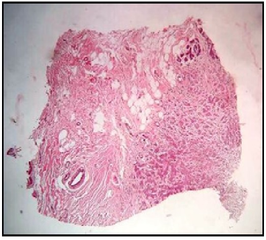
Materials and Methods: Tissue cores were obtained from random tissues which included placenta, breast tissue and lymphnode and endometrium using skin punch biopsy needle of bore 2 mm. These were done for standardizing the procedure of a miniature tissue microarray. Further, tissue cores obtained from carcinoma breast tissue by two different methods were used for constructing a microarray block. Sections of 4 micron thickness were taken and stained with hematoxylin and eosin stains. If satisfactory tumor tissue is present in this constructed block, then sections were taken for immunohistochemistry staining with ER, PR and HER2.
Results: Directly constructed tissue blocks had better preservation of tumor tissue morphology compared to the blocks constructed from donor blocks. Also, directly constructed tissue blocks had the advantage of not mutilating the donor block which could be still used for further studies and reference. Immunohistochemistry revealed similar results as obtained during the routine histopathological sections. Also, the cost of the reagents used for immunohistochemistry was reduced by 200% as compared to the routine immunohistochemical staining procedure.
Conclusion: A petit Tissue microarray is definitely possible in a tertiary care histopathology laboratory and can be utilized for immunohistochemical studies with multiple markers.
Downloads
References
2. Kallioniemi OP, Wagner U, Kononen J, Sauter G. Tissue microarray technology for high-throughput molecular profiling of cancer. Hum Mol Genet. 2001 Apr;10(7):657-62.[pubmed]
3. Avninder S, Ylaya K, Hewitt S. Tissue microarray: A simple technology that has revolutionized research in pathology. Journal of Postgraduate Medicine. 2008;54(2):158.Doi.10.4103/0022-3859.40790
4. Choi C, Kim K, Song J, Choi S, Kim L, Park I et al. Construction of High-Density Tissue Microarrays at Low Cost by Using Self-Made Manual Microarray Kits and Recipient Paraffin Blocks. Korean Journal of Pathology. 2012;46(6):562.DOI.10.4132/KoreanJPathol.2012.46.6.562
5. Shebl A, Zalata K, Amin M, El-Hawary A. An inexpensive method of small paraffin tissue microarrays using mechanical pencil tips. Diagnostic Pathology. 2011;6(1):117.DOI.org/10.1186/1746-1596-6-1176.
6. Tripodi SA, Rocca BJ, Hako L, Barbagli L, Bartolommei S, Ambrosio MR. Quality control by tissue microarray in immunohistochemistry. Journal of Clinical Pathology. 2012;Jul;65(7):635-7. doi: 10.1136/jclinpath-2011-200551.
7. Gulbahce H, Gamez R, Dvorak L, Forster C, Varghese L. Concordance Between Tissue Microarray and Whole-section Estrogen Receptor Expression and Intratumoral Heterogeneity. Applied Immunohistochemistry & Molecular Morphology. 2012;20(4):340-343.doi: 10.1097/PAI.0b013e318241ca14
8. Mengel M, Kreipe H, von Wasielewski R. Rapid and Large-Scale Transition of New Tumor Biomarkers to Clinical Biopsy Material by Innovative Tissue Microarray Systems. Applied Immunohistochemistry & Molecular Morphology. 2003;11(3)261-268.
9. Matysiak B, Brodzeller T, Buck S, French A, Counts C, Boorsma B et al. Simple, Inexpensive Method for Automating Tissue Microarray Production Provides Enhanced Microarray Reproducibility. Applied Immunohistochemistry & Molecular Morphology. 2003;11(3)269-273.[pubmed]
10. Chavan SS, Ravindra S, Prasad M. Breast Biomarkers-Comparison on Whole Section and Tissue Microarray Section. Journal of Clinical and Diagnostic Research : JCDR. 2017;11(3):EC40-EC44. doi:10.7860/JCDR/2017/25088.9573.
11. Srinath S, Kendole RK, Gopinath P, Krishnappa S, Vishwanath S. Economic methods used in fabrication of tissue microarray: A pilot study. Journal of Oral and Maxillofacial Pathology : JOMFP. 2016;20(1):86-90. doi:10.4103/0973-029X.180948.
12. Bhargava R, Lal P, Chen B. Feasibility of using tissue microarrays for the assessment of HER-2 gene amplification by fluorescence in situ hybridization in breast carcinoma. Diagn Mol Pathol. 2004 Dec;13(4):213-6.

 OAI - Open Archives Initiative