
Comparative study using routine stains and Immunohistochemistry staining techniques for detection of Helicobacter pylori
Abstract
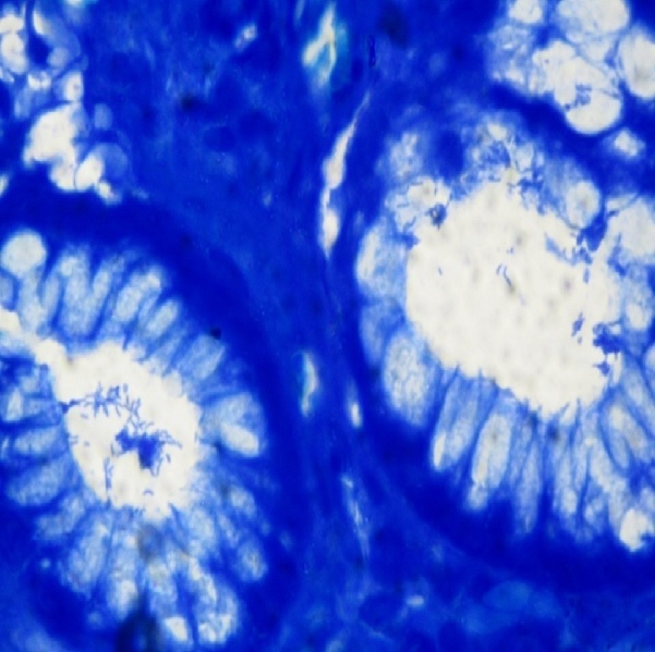
Introduction: The aim of this study was to evaluate the role of endoscopic biopsies for detection of Helicobacter Pylori using routine staining techniques which included hematoxyline and eosin, Giemsa and Immunohistochemistry.
Methods: A prospective study of 3 years in which 53 gastric endoscopic mucosal biopsies were included. These patients had a clinical history of dyspeptic symptoms. 3 staining techniques were used to identify H. Pylori. Hematoxylin and eosin staining was done routinely along with Giemsa and H. Pylori detection using antibodies directed against specific antigens in IHC. The staining pattern of H. Pylori was also studied.
Results: In 53 cases studied, modified Giemsa staining is the cheapest and easiest to perform but antibodies directed against specific antigens in IHC proved to be more specific in identifying H. Pylori than other staining techniques. While Giemsa staining was non-specific for other species of Helicobacter IHC was specific for H. Pylori. Spiral type of staining was the most frequent of the staining pattern.
Conclusion: Our study highlighted the association of Helicobacter Pylori in patients with functional dyspepsia and proved Immunohistochemistry as gold standard in identifying Helicobacter Pylori with Geimsa being practically applicable in Indian set up keeping the cost factor in mind.
Downloads
References
2. Dunn BE , Cohen H, Blaser MJ. Helicobacter pylori. Clin Microbiol Rev. 1997 Oct;10(4):720-41.[pubmed]
3. Kusters JG , Gerrits MM, Van Strijp JA, Vandenbroucke-Grauls CM. Coccoid forms of Helicobacter pylori are the morphologic manifestation of cell death. Infect Immun. 1997 Sep;65(9):3672-9.[pubmed]
4. Anim JT , Al-Sobkie N, Prasad A, et al. Assessment of different methods for staining Helicobacter pylori in endoscopic gastric biopsies. DOI:10.1078/S0065-1281(04)70022-7.
5. Sulakshana MS, Siddiq M, Ahmed Raghupathi AR. A histopathological study of association of Helicobacter pylori with gastric Malignancies, Int. J Curr Res Aca Rev. 2015; 3(3):10-28.
6. Schistosomes, liver flukes and Helicobacter pylori. IARC Working Group on the Evaluation of Carcinogenic Risks to Humans. Lyon, 7-14 June 1994. IARC Monogr Eval Carcinog Risks Hum. 1994;61:1-241.[pubmed]
7. Moayyedi P , Dixon MF. Any role left for invasive tests? Histology in clinical practice. Gut. 1998 Jul;43 Suppl 1:S51-5.[pubmed]
8. Doglioni C , Turrin M, Macrì E, et al. HpSS: a new silver staining method for Helicobacter pylori. J Clin Pathol. 1997 Jun;50(6):461-4.[pubmed]
9. Rotimi O , Cairns A, Gray S,v et al. Histological identification of Helicobacter pylori: comparison of staining methods. J Clin Pathol. 2000 Oct;53(10):756-9.[pubmed]
10. Dixon MF , Genta RM, Yardley JH, Correa P. Classification and grading of gastritis. The updated Sydney System. International Workshop on the Histopathology of Gastritis, Houston 1994. Am J Surg Pathol. 1996 Oct;20(10):1161-81.[pubmed]
11. Wabinga HR. Comparison of immunohistochemical and modified Giemsa stains for demonstration of Helicobacter pylori infection in an African population. Afr Health Sci. 2002 Aug;2(2):52-5.[pubmed]
12. Ashton-Key M, Diss TC, Isaacson PG. Detection of Helicobacter pylori in gastric biopsy and resection specimens. J Clin Pathol. 1996 Feb;49(2):107-11.[pubmed]
13. Lee JY , Kim N . Diagnosis of Helicobacter pylori by invasive test: histology. 10.3978/j.issn.2305-5839.2014.11.03.[pubmed]
14. Histological identification of H. pylori stained by hematoxylin-eosin and Giemsa: review for quality control J Bras Patol Med Lab, v. 51, n. 2, p. 108-112, April 2015 Marcela S. Boldt¹; Rivelle D. Pereira¹; Alfredo J. A. Barbosa²
15. Rosai J. Gastrointestinal tract-Stomach. Rosai and Ackerman s Surgical Pathology. Missouri: 9th ed. Elsevier Inc; 2009: 650- 684.
15. Yodavudh S, Tangjitgamol S, Puangsa S. Mixture of carbol fuchsin and alcian blue staining of gastric tissue for the identification of H.pylori and goblet cell intestinal metaplasia. Southeast Asian J Top Med Pub health; Jul 2008, 39 (4): 656-666.
16. Soylu A, Ozkara S, Alıs H, et al. Immunohistochemical testing for Helicobacter Pylori existence in neoplasms of the colon. BMC Gastroenterology. 2008;8:35. doi:10.1186/1471-230X-8-35.Riba AK, Ingeneri TJ, Strand CL. Improved histologic identification of Helicobacter pylori by immunohistochemistry using a new Novocastra monoclonal antibody. Lab Med [Internet]. 2011;42(1):35–9.
17. Jhala NC , Siegal GP, Klemm K, et al. Infiltration of Helicobacter pylori in the gastric mucosa. DOI: 10.1309/YDTX-KE06-XHTH-FNP2. [pubmed]
18. Patnayak K, Reddy V, Jena A, Rukmangadha N, Panhasarathy S, Reddy MK. Utility of immunohistochemistry in demonstrating Helicobacter pylori. Oncol Gastroenterol Hepatol. 2015;4:4–7.

 OAI - Open Archives Initiative