
Correlation of coronary artery atherosclerosis typing and luminal narrowing with ischemic myocardial lesions in post mortem heart specimens: a four year retrospective study
Abstract
Introduction: Atherosclerosis accounts for a large proportion of cardiovascular system associated morbidity and mortality. Coronary artery disease (CAD) is the leading cause of global deaths with about 80% of burden occurring in developing countries.
Material & Methods: In order to assess the magnitude of the problem, a retrospective study of autopsy cases for the presence of atherosclerotic lesions of coronary arteries and associated ischemic cardiac lesionswas under taken from January 2013 to December 2017. Also, correlation of the atherosclerosis with ischemic heart diseases was studied.
Result: Total number of heart specimens received in department of Pathology during four years were 272. Out of these 57 were autolyzed and were excluded from the study. Significant atherosclerotic lesions were seen in 54(25.11%) and 64(29.76%) cases in right and left coronary arteries respectively and were statistically significantly higher among age group > 41 years as compared to those with age <41 years, overall atherosclerotic lesions were significantly higher in age groups >41 years as compared to <41 years.
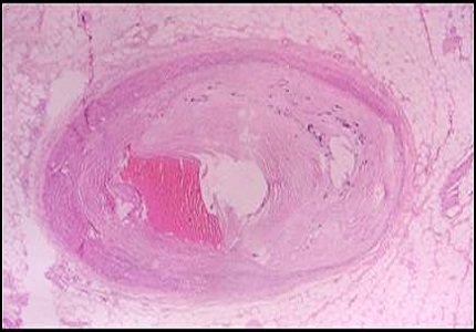
Conclusion: Maximum number of significant cardiac lesion were associated with atherosclerotic type VIII lesions (75%) followed by type VII (66.66%) and type VI (33.33%). Maximum number of significant myocardial lesions were associated with grade IV (66.66%) coronary luminal narrowing followed by grade III (45.71) and grade II (38.46). The study also showed significant correlation between the higher grade of the coronary atherosclerotic lesions and the ischemic heart disease.
Downloads
References
2. Indrayan A. Forecasting vascular disease cases and associated mortality in India. NCMH Background Papers: Burden of Disease in India. National commission on macroeconomics and Health, Government of India; 2005.p.197-215.
3. Gaziano TA. Cardiovascular disease in the developing world and its cost-effective management. Circulation. 2005; 112(23):3547-53.doi:10.1161/CIRCULATIONAHA.105.591792.
4. Beaglehole R, Reddy S, Leeder S. Poverty and human development. The global implications of cardiovascular disease. Circulation. 2007; 116:1871-1873. [PubMed]
5. Bertomeu A, García-Vidal O, Farré X, Galobart A. Preclinical coronary atherosclerosis in a population with low incidence of myocardial infarction: cross sectional autopsy study. BMJ. 2003; 327:591- 2.
6. Stary HC. Natural History and Histological Classification of Atherosclerotic Lesions: An Update. ArteriosclerThrombVasc Biol. 2000; 20; 1177-1178. [PubMed]
7. Porwal V, Khandelwal S, Jain D, Gupta S.Histological Classification of Atherosclerosis and Correlation with Ischemic Heart Disease: A Autopsy Based Study. Annals of Pathology and Laboratory Medicine. 2016; 3(2):99-104.
8. Bavelaar FJ, Beynen AC. Atherosclerosis in parrots: a review. Vet Q. 2004;26:50–60.
9. Brunkwall J, Mattsson E, Bergqvist D. Dietinducedatherosclerosis in rabbits alters vascularprostacyclin release. Eicosanoids. 1992;5:197–202.
10. Fricke C, Schmidt V, Cramer K. Characterization of atherosclerosis by histochemical andimmunohistochemical methods in African grey parrots (Psittacus erithacus) and Amazon parrots(Amazona spp.). Avian Dis. 009;53:466–72. [PubMed]
11. Centers for Disease Control and Prevention CDC. Racial/ethnic and socioeconomic disparities inmultiple risk factors for heart disease and stroke. MMWR Morb Mortal Wkly Rep. 2005;54:113–7.
12. Galkina E, Ley K. Immune and inflammatory mechanisms of atherosclerosis. Annu Rev Immunol.2009;27:165–97.
13. Hansson GK, Libby P. The immune response in atherosclerosis: a doubleedgedsword. Nat Rev Immunol. 2006;6:508–19. [PubMed]
14. Silverman S. Diagnostic Imaging. In: Mader DR, editor. Reptile Medicine and Surgery. 2nd ed. St.Louis: Saunders Elsevier; 2006. p. 471489.
15. Willecke F, Yuan C, Oka K, Chan L, Hu Y, Barnhart S, et al. Effects of High Fat Feeding and Diabetes on Regression of Atherosclerosis Induced by LowDensityLipoprotein Receptor Gene Therapy in LDL ReceptorDeficientMice. PLoS One. 2015;10 (6):e0128996.
16. Smith SC, Smith EC, Taylor Jr RL. Susceptibility to spontaneous atherosclerosis in pigeons: anautosomal recessive trait.J. Heredity. 2001;92:439–42.
17. Bairey NCB, Merz BD, Johnson BL, Sharaf WISE. Hypoestrogenemia of hypothalamic origin and coronary artery disease in premenopausal women: a report from the NHLBIsponsored WISE study. J Am Coll Cardiol. 2003;41:413–9. [PubMed]
18. Kumar V, Abbas A, Aster J. The Heart. Robbins And Cortans Pathological Basis Of Disease. 2015; 9th Ed:523-578.
19. Murthy MSN, Dutta BN, Ramalingaswami V. Coronary atherosclerosis in North India (Delhi Area). J PatholBacteriol.1963;85:93-101.
20. Singh H, Oberoi SS, Gorea RK, Bal MS. Atherosclerosis in Coronaries in Malwa Region of Punjab. J Indian Acad Forensic Med. 2005;27(4):32-5.
21. Padmavati S, Sandhu I. Incidence of coronary artery disease in Delhi from medico-legal autopsies. Indian Journal of Medical Research. 1969; 57:465-475.
22. Tandon OP, Aggarwal VC, Katiyar BC. Coronary and aortic atherosclerosis. Indian Heart J.1969;5:10.
23. Bhargava MK, Bhargava SK. Coronary atherosclerosis in North Karnataka. Indian J Pathol Microbiol. 1975;18:65-77. [PubMed]
24. Chirinos JA, Segers P, Duprez DA, Brumback L, Bluemke DA, Zamani P, et al. Late systolic central hypertension as a predictor of incident heart failure: the Multiethnic Study of Atherosclerosis. J Am Heart Assoc. 2015;4 (3):e001335.
25. Bertomeu A, GarcíaVidal O, Farré X. Preclinical coronary atherosclerosis in a population with low incidence of myocardial infarction: cross sectional autopsy study. British Med J. 2003;327(7415):591–2. [PubMed]
26. Dhruva GA, Agravat AH, Sanghvi HK. Atherosclerosis of coronary arteries as predisposing factor in myocardial infarction: An autopsy study. Online J Health Allied Scs. 2012 11:1.
27. Stetter MD. Ultrasonography. In: Mader DR, editor. Reptile Medicine and Surgery. 2nd ed. St. Louis: Saunders Elsevier; 2006. p. 6656-74.
28. Pearson TA, Blair SN, Daniels SR, Eckel RH, Fair JM, Fortmann SP. AHA guidelines for primary prevention of cardiovascular disease and stroke: 2002 update: Consensus panel guide to comprehensive risk reduction for adult patients without coronary or other atherosclerotic vascular diseases. American Heart Association Science Advisory and Coordinating Committee. Circulation.2002;106:388–91. [PubMed]
29. Widimsky P, Andel M. Prevalence of coronary atherosclerosis in asymptomatic population. Eur Heart J.2000;21:13-14.
30. Yazdi SAT, Rezaei A, Azari JB, Hejazi A, Shakeri MT, Shahri MK. Prevalence of Atherosclerotic Plaques in Autopsy Cases with Noncardiac Death. Iranian J Pathol. 2009;4(3):101-104.
31. Golshahi J, Rojabi P, Golshahi F. Frequency of atherosclerotic lesions in coronary arteries of autopsy specimens in Isfahan forensic medicine center. J Res Med. 2005;1(10):16-9.
32. Wig KL, Malhotra RP, Chitkara NL, Gupta SP. Prevalence of Coronary Atherosclerosis In Northern India. BMJ.1962;2:510-3. [PubMed]
33. Sudha ML, Sundaram S, Purushothaman KR, Kumar PS, Prathiba D. Coronary atherosclerosis in sudden cardiac death: An autopsy study. Indian J Pathol Microbiol. 2009;52(4):486-9.
34. Virmani R, Kolodgie FD, Burke AP, Farb A, Schwartz SM. Lessons from sudden coronary death – A comprehensive morphological classification scheme for atherosclerotic lesions. ArteriosclerThrombVasc Biol.2000;20:1262-75.
35. Stary HC, Chandler AB, Glagov S, Guyton JR, InsullW,Rosenfeld ME, Schaffer A, Schwartz CJ, Wagner WD, Wissler RW. A definition of initial, fatty streak, and intermediate lesions of atherosclerosis. ArteriosclerThromb. 1994;14:840–856. [PubMed]
36. Shirani J, Youseti, Roberts WC. Major cardiac findings at necropsy in 366 American octogenarians. Am J Cardiol. 1995:75,151-156.
37. Mcgill HC, Brown BW, Gorel et al. Grading stenosis in the right coronary artery. Circulation. 1968; 37:460-468. [PubMed]
38. Maru M. Coronary atherosclerosis and myocardial infarction in autopsied patients in Gondar, Ethiopia. J R Soc Med.1989;82(7):399-40. [PubMed]
39. Bharti Jha et al. Incidence of coronary atherosclerosis in different coronary arteries and its relation with myocardial infarction: a randomized study in 300 autopsy heart in tertiary care hospital.International journal of Medical Science and Public Health. 2013; 2: 836-839.
40. Shiladeria P et al.Coronary atherosclerosis and myocardial infarction, an autopsy study. NJIRM. 2013;4: 106-108. [PubMed]

 OAI - Open Archives Initiative