
Comparison of Z. N. staining & fluorescent microscopy in detection of M. Tuberculosis bacilli in Fine needle aspiration smears
Abstract
Aims: To compare ZN stain method with Fluorescent method for detection of tuberculous bacilli in FNA smears in terms of sensitivity & feasibility.
Settings and Design: A Prospective study was conducted in the department of Pathology at tertiary care center. FNAC done from lymph node lesion in clinically suspected cases of tuberculosis attending the Department of Medicine, Surgery, ENT, TB and Chest.
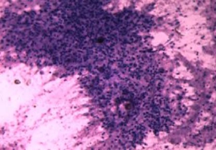
Methods and Material: Fine-needle aspiration was performed in the Department of Pathology from January 2016 to February 2017. Out of 409 overall FNAC samples, there are 193 FNAC Lymph nodelesions, out of them 65 clinically suspected cases were processed for direct microscopy using conventional ZN staining and routine cytology and compared with the findings of the modified fluorescent method.
Statistical analysis used: Simple statistical analysis done by using χ2 test.
Results: Out of the 65 Tuberculous positive aspirates, the smear positivity for AFB on the ZN method was 43.07% (28/65) while the positivity increased to 87.69% (57/65) on the Auramine-Rhodamine fluorescent method.
Conclusions: Fluorescent microscopy is rapid, simple, easy method with high detection rate for AFB as compared to ZN method.
Downloads
References
2. Lau SK, Wan SK, and Wei WI, “Source of tubercle bacilli in cervical lymph nodes: a prospective study, J LaryngolOtol1991;105:558-61.
3. PahwaRD,Jain et al Assessment of possible tuberculous lymphadenopathy, by PCR compared to non-molecular methods, J Med Microbio2005;54:873-78. [PubMed]
4. Singh KK, Murlidhar M, Kumar et al, Comparison of in house polymerase chain reaction with conventional techniques for the detection of Mycobacterium tuberculosis DNA in granulomatous lymphadenopathy, J ClinPathol 2000;53:355-61. [PubMed]
5. AnnamV, Kulkarni MH and Puranik RB, Comparison of the modified fluorescent method and conventional ZiehlNeelsen method in the detection of acidfast bacilli in lymphnode aspirates, J cytol 2009;6:40-43.
6. Jain A, Bhargava A, and Agarwal SK, A comparative study of two commonly used staining techniques for acid fast bacilli in clinical specimens.Indian J Tuberc.2002;49:161-62.
7. WrightCA,BurgessSM,BlumbergL. Mycobacterialautofluorescence in Papanicolaou stained lymph node aspirates: a glimmer in the dark. DiagnCytopathol2004;30:257-60.
8. C. A. Wright, A. C. Hesseling, C. Bamford, S. M. Burgess, R. Warren, and B. J. Marais, “Fine-needle aspiration biopsy: a first-line diagnostic procedure in paediatrictuberculosis suspects with peripheral lymphadenopathy?” International Journal of Tuberculosis and Lung Disease, 2009; 13(11):1373-79
9. Holst E, Mitchison DA, Radhakrishna S. Examination of smears for tubercle bacilli by fluorescent microscopy. Indina J Med Res 1959;47:495-9. [PubMed]
10. Sommers HM, Good RC. Mycobacterium. In: Lennette EH, Balows A, Hausler WJ, Shadomy HJ, editor. Manual of Clinical Microbiology. 4th ed. American Society for Microbiology. Washington.D.C: 1985: 216-48.
11. Winn WC, Allen SD, Janda WM, Koneman EW, Procop GW, Schreckenberger, et al. In: Mycobacteria. In Koneman’s Color atlas and text book of diagnostic microbiology. 6th ed. Philadelphia: Lippincott Williams and Wilkins; 2006:1071-72.
12. V.S.RajanandY.S.Goh. Intermittent chemotherapy in the treatment of tuberculosis cutis. A preliminary report. British Journal of Dermatology. 1972;87(3):270–273.
13. Daniel TM, “The rapid diagnosis of tuberculosis: a selective review,” J Lab Clin Med 1990;116: 277-82. [PubMed]
14. Dagar et al, Comparision of ZN Staining and Fluorescent Microscopy in Detection of Acid Fast Bacilli in Fine Needle Aspiration Smears IOSR JDMS2016;15:79-84
15. Annam V, Kulkarni MH, Puranik RB. Comparison of the modified fluorescent method and conventional Ziehl–Neelsen method in the detection of acid-fast bacilli in lymphnode aspirates J cytol2009;6:13. [PubMed]
16. Thakur B, Mehrotra R, Nigam JS. Correlation of various techniques in diagnosis of tuberculous lymphadenitis on fine needle aspiration cytology. Patholog Res Int 2013: 2013: 824620. [PubMed]

 OAI - Open Archives Initiative