
Umbilical mucosal polyp in an infant: a rare entity Mimicking umbilical granuloma
Abstract
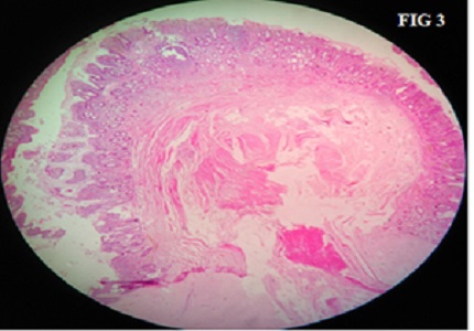
Umbilical lesions constitute a comparatively lesser percentage of the specimens received on a daily basis for histopathological examination. During embryological development, the umbilicus functions as a channel that allows flow of blood between the placenta and fetus. It also serves an important role in the development of the intestine and the urinary system. After birth, once the umbilical cord falls off, no evidence of these connections should be present. Nevertheless a few lesions are encountered related to the same. Patients with umbilical disorders present with drainage, a mass, or both. Umbilical granuloma, omphalomesenteric remnants, urachal remnants, hernias are few lesions in the umbilical region. We present a case of an umbilical polyp in an infant, which clinically suspected to an umbilical granuloma. The idea behind presenting this case was that, not only are umbilical polyps rare lesions but also it is necessary to differentiate it from umbilical granuloma as the treatments may vary. The clinicians and reporting pathologists must be aware of this rare congenital lesion.
Downloads
References
2. Gaopande VL, Deshmukh SD, Khandeparkar SS, Suryavanshi MA, Patil VR. Clinical and histopatho-logical profile of lesions of umbilicus. Medical Journal Of DY Patil University. 2015;8(2):179-81. doi: 10.4103 / 0975-2870.153152.
3. Pacilli M, Sebire NJ, Maritsi D, Kiely EM, Drake DP, Curry JI, et al. Umbilical polyp in infants and children. Eur J Pediatr Surg. 2007 Dec;17(6):397-9. [PubMed]
4. Pomeranz A. Anomalies, Abnormalities and Care of the umbilicus. Pediatr Clin North Am. 2004 Jun;51 (3): 819-27, xii.
5. Steck WD, Helwig EB. Cutaneous remnants of the omphalomesenteric duct. Arch Dermatol. 1964 Nov;90: 463-70. [PubMed]
6. Karagüzel G, Aldemir H. Umbilical Granuloma: Modern understanding of etiopathogenesis, diagnosis, and management. J Pediatr Neonatal Care. 2016; 4(3): 00136. doi: 10.15406/jpnc.2016.04.00136.
7. You Y, Yang X, Hao F, Zhong B. The umbilical polyp: A report of two cases and literature review.Int J Dermatol. 2009 Jun; 48 (6):630-2. doi: 10.1111/j.1365-4632. 2009.03880.x.
8. Brady M, Conway AB, Zaenglein AL, Helm KF. Umbilical Granuloma in a 2 month old patient: Histo-pathology of a common clinical entity. Am J Dermatopathol. 2016 Feb; 38 (2):133-4. doi:10.1097/ DAD. 0000000000000429. [PubMed]
9. Piparsaliya S, Joshi M, Rajput N, Zade P. Patent Vitellointestinal Duct: A close differential diagnosis of umbilical granuloma: A case report and review of literature. Surgical Science. 2011;2(3):134-6 doi:10. 4236/ss.2011.23027.

 OAI - Open Archives Initiative