
Histopathological study of skin tumours
Abstract
Background: Skin tumour and their various histological typesalways creating a diagnostic difficulty. Histopathology is the gold standard for diagnosis in turn is used by clinicians to aid clinical outcome andalso emphasizing certain key forvaluable observation of certain tumours.
Objectives: The study was conducted to find out the occurrence of different tumours of skin in MGMCRI and to classify skin tumours (WHO classification).
Methods: The pathological features of 52 cases of Skin Tumours were analysed between May 2009 and May 2014. Section of skin tissue was studied by light microscopy after hematoxylin and eosin staining.
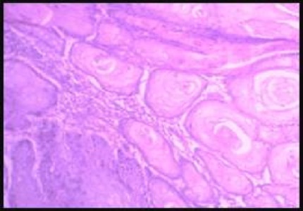
Results: A total number of 52 cases of skin tumours were studied. Benign skin tumours constituted 44.23% while malignant skin tumours constituted 55.76% of all skin tumours. Intradermal nevus was the most common benign Skin tumours while Squamous Cell Carcinoma was the most common malignant tumours.
Conclusion: Light Microscopic examination remains the standard technique for the diagnosis of the tumours. Although special stain and immunochemistry can be utilised in situation where the diagnosis cannot be confirmed on hematoxylin and eosin.
Downloads
References
2. Gundalli S, Kolekar R, Pai K, Kolekar A. Histopathological Study of Skin Tumours. International Journal of Healthcare Sciences.2015 March; 2(2): 155-163.
3. Weinstock MA. Death from skin cancer among the elderly: epidemiological patterns. Arch Dermatol.1997. 133(10): 1207-1209. [PubMed]
4. McCourt C, Dolan O, Gormley G. Malignant melanoma: a pictorialreview. Ulster Med J.2014 May;83(2):103-10. [PubMed]
5. Tirumalae R, Roopa MO. Beningvs Malignant skin adnexal Neoplasms: How useful are silhouettes? Indian Journal of Dermatology. 2013Jan; 58(1):30-33.doi: 10.4103/0019-5154.105282. [PubMed]
6. Kempf W, Hantschke M, Kutzner H, Burgdorf W. Dermatopathology. adnexal tumour Germany Springer 2008.
7. Roncalli De oliveria W, Neto CF, Rady PL, Tyring SK. Seborrheic keratosis – like lesions in patients with epidermodysplasia verruciformis. J Dermatol. 2003 Jan;30(1): 48-53.
8. Sanjay N Parate, Rishi B Chahande, Suprita P Nayak and Sudhakar K Bobhate. Adnexal tumours of skin. Ind J Dermat.1998June ;43(2):58-60.
9. LeBoit PE, Burg G, Weedon D, Sarasin A. World Health Organization Classification of Tumours Pathology and Genetics of Skin Tumours.3rd Edition. France: IARC Press, International Agency for Research on Cancer,150 cours Albert Thomas, F-69008 Lyon, France ;2006.
10. Shaikh SS. Histopathological study of skin tumours ( M.D thesis). Hubli Karnataka :2011. [PubMed]
11. Baruah B, Sengupta, Kesari SP, Ilapakurty B. Pattern on Non-Melanoma skin Cancer in Sikkim, India: 13-years clinicopathological review. Indian J Otolaryngol Head Neck Surg.2013July ;65(suppl1):160-162.
12. Deo SV, Sidhartha H, Shukla N K, Kumar S, Kar M, Samaiya A. Surgical management of skin cancers: Experience from a regional cancer centre in north India. Indian journal of cancer .2005 September ;42(3):145-50.
13. Alakloby OM, Bukhari I A, Shawarby M.A. Histopathological pattern of Non –melanoma skin cancer at King Fahd Hospital of the university in the Eastern region of Saudi Arabia during the years 1983 to 2002.Cancer therapy.2008 May ;6(1) :303-306.
14. Radhika K, Phaneendra BV, Rukmangadha N, Reddy MK. A study of biopsy confirmed skin adnexal tumours:experience at a tertiary care teaching hospital.The Journal of Clinical and Scientific Research.2013 July ;2(3):132-8.
15. Sharma A, Paricharak DG, Nigam JS, Rewri S, Soni PB, Omhare A, Sekar P. Histopathological study of Skin Adnexal Tumours- Institutional Study in South India. Journal of Skin cancer.2014;1-4. [PubMed]
16. Kaur K, Gupta K, Hemrajani D, Yadav A, Mangal K. Histopathological analysis of skin adnexal tumors: A three year study of 110 cases at a tertiary care center. Indian J Dermatol. 2017Aug;62(4):400-6. [PubMed]
17. Paudyal P, Agarwal M, Pradhan A, Sinha AK, Agrawal S.A clinico-histopathological study on skin appendageal tumors. Journal of Pathology of Nepal2016 ; 6(11) : 885-91.
18. Deka M, Bhattacharjee S. Histopathology of Skin Adnexal Tumors- A Two Year Retrospective Study at a Tertiary Care Hospital. Int J Sci Stud .2016 Dec;4(9):61-65.

 OAI - Open Archives Initiative