
Histopathological analysis of hysterectomy specimens in tertiary care center: two year study
Abstract
Background: The uterus is a major female hormone-responsive reproductive pear-shaped organ located within the pelvic region. The gross morphology of uterus quite dramatically changed throughout life by changing levels of ovarian hormones. A hysterectomy is done for many reasons including the ridding of tumours both benign and malignant. It is the most commonly performed gynecological surgical procedure throughout the world.
Materials and Methods: This study was undertaken to identify the different types of pathologies in hysterectomy specimens and to correlate the findings with the different age group and clinical indications.In the present study, 303 cases were studied over a period of two years from January 2016 to December 2017. Surgical specimens were formalin fixed and the tissue was adequately processed for histopathological examination. The sections were stained routinely with hematoxylin and eosin stain.
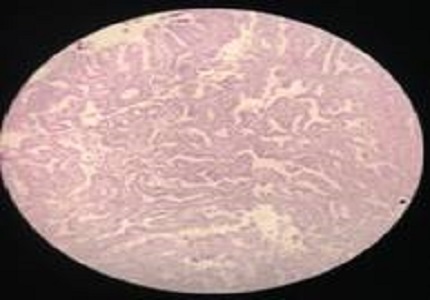
Results: Of the 303 cases, half of the cases (50.8%) were encountered in the age group of 40 - 49 years which was the most common age group. Fibroid uterus (38.6%) and uterine prolapse (32.6%) were the most common clinical indications for hysterectomy. The most common pathology identified was leiomyoma (42.2%) in myometrium. In cervix most common finding was chronic cervicitis (28.1%). Histopathological examination confirmed the clinical and gross diagnosis in majority of the cases.
Conclusions: The present study provides a fair insight into the histological patterns of lesions in hysterectomy specimens in our institution. A wide range of lesions are encountered when hysterectomy specimens are subjected to histopathological examination. Few lesions were encountered as incidental findings. Hence, it is important that every hysterectomy specimen should be subjected to detailed gross and histopathological examination for better postoperative management.
Downloads
References
2. Nausheen F, Iqbal J, Bhatti FA, Khan AT, Sheikh S. Hysterectomy: the patient's perspective. Annals of King Edward Medical University. 2016 May;10(4):339-41.
3. Silverberg SG, DeLellis RA, Frable WJ, LiVolsi VA, Wick MR. Silverberg’s Principles and practice of surgical pathology and cytopathology.4th ed. Eddinburg: Elsevier;2006.Vol.2.1935.
4. Jaleel R, Khan A, Soomro N. Clinicopathological study of abdominal hysterectomies. Pak J Med Sci.2009;25(4):630-34.
5. Watts WF,Kimbrough RA Jr. Hysterectomy; analysis of 1000 consecutive operations. Obstet Gynecol. 1956 May;7(5):483-93. [PubMed]
6. Rather GR, Gupta Y, Bardhwaj S. Patterns of Lesions in Hysterectomy Specimens: A prospective study. JK Sci J Med Edu Res. 2013;15(2):63-68.
7. Ramachandran T, Sinha P, Subramanium. Correlation between Clinicopathological and Ultrasonographical Findings in Hysterectomy. Journal of Clinical and Diagnostic Research. 2011;5(4):737-740.
8. Ajmera sachin K, Mettler L, and Jonat W. Operative spectrum of hysterectomy in a German university hospital. J ObstetGynecol India. 2006;56(1):59-63.
9. Chandralekha J, Sumanlatha GR, Kartheek BVS, Bhagyalakshmi A. Prospective study of uterine corpus lesions over a period of one year in tertiary care centre. Int J Res Med Sci.2016;4:2583-7.
10. Raza AKMM. Histological Findings in Hysterectomy Specimens in a Tertiary Medical College Hospital in Bangladesh. J CytolHistol 2017;2(1):3.
11. Dhuliya V, Gosai D, Jain H, Goswami H. Histopathological study of uterine and cervical lesion in hysterectomy specimen. BJkines-NJBAS. 2016;8(2):23-26.
12. Baral R, Sherpa P, Gautam D.Histopathological analysis of hysterectomy specimens: one year study. Journal of Pathology of Nepal. 2017;7:1084-1086.
13. Emons G, Beckmann MW, Schmidt D, Mallmann P. New WHO Classification of Endometrial Hyperplasias. Uterus commission of the Gynecological Oncology Working Group (AGO), GeburtshilfeFrauenheilkd. 2015;75(2):135.
14. Ranabhat SK. Shrestha R. Tiwari M, Sinha DP. Subedee LR. A retrospective histopathological study of Hysterectomy with or without salpingo-ophorectomy specimens. JCMC. 2010;1(1):26-29.
15. Ojeda VJ. The pathology of hysterectomyspecimens.N Z Med J. 1979 Mar 14;89(631):169-71. [PubMed]
16. Abdullah LS. Hysterectomy: A Clinicopathologic Correlation. Bahrain Medical Bulletin. 2006 June;28(2): 1-6.
17. Mehboob R, Ahmad N. Unexpected pathology at vaginal hysterectomy for genital prolapse. Pak J Med Res2002;41(4):142-44.
18. Talukder SI, Haque MA, Huq MH, Alam MO, Roushan A, Noor Z, Nahar K. Histopathologicalanalysis of hysterectomyspecimens.Mymensingh Med J. 2007 Jan;16(1):81-4. [PubMed]

 OAI - Open Archives Initiative