
Intraorbital, extraconal cavernous hemangioma: a common tumor at an uncommon site-a case report
Abstract
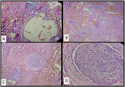
A wide variety of processes can produce space occupying lesions in and around the orbit including benign neoplasms, malignant neoplasms, vascular lesions, inflammatory diseases, congenital lesions etc. Cavernous hemangioma is the most common benign non-infiltrative neoplasm of the orbit. It is also the most common intraorbital tumor found in adults.Although histologically benign, they can encroach on intraorbital or adjacent structures and be considered anatomically or positionally malignant. We present here a case of extraconal, intraorbital cavernous hemangioma in a 21 years old male diagnosed histopathologically. So, we conclude that even though intraorbital cavernous hemangiomas present most frequently at intraconal locations, extraconal site for its occurrence cannot be ignored and must be considered in evaluation of intraorbital vascular lesions.
Downloads
References
2. Anand R, Deria K, Sharma P, Narula MK, and Garg R. Extraconal cavernous hemangioma of orbit: A case report. Indian J Radiol Imaging. 2008 Nov; 18(4): 310–312.
3. Rosai J, editor. Rosai and Ackerman’s Surgical Pathology. 10th ed. Edinburgh: Elsevier; 2011.
4. Weiss SW, Goldblum JR, editors. Enzinger and Weiss's Soft Tissue Tumors, 5th Ed. Mosby: Elsevier; 2008.
5. Stamm A, Nogueira JF. Orbital cavernous hemangioma: Transnasal endoscopic Management. Otolaryngology–Head and Neck Surgery 2009; 141, 794-795.
6. Wilms G, Raat H, Dom R, Thywissen C, Demaerel P, Dralands G, Baert AL. Orbital cavernous hemangioma: findings on sequential Gd-enhanced MRI. J Comput Assist Tomogr. 1995 Jul-Aug;19(4):548-51.
7. Bilaniuk LT. Orbital vascular lesions. Role of imaging. Radiol Clin North Am. 1999 Jan;37(1):169-83, xi.
8. Harris GJ, Jakobiec FA. Cavernous hemangioma of the orbit. J Neurosurg. 1979 Aug;51(2):219-28. [PubMed]
9. Zenobii M, Galzio RJ, Lucantoni D, Caffagni E, Magliani V.Spontaneousintraorbitalhemorrhagecaused bycavernous angioma of the orbit. J Neurosurg Sci. 1984 Jan-Mar;28(1):37-40. [PubMed]
10. Rizvi S, Yousuf S, Maheshwari V, Khan R. Multiple cavernous haemangiomas of the the orbit and conjunctiva: Arare association. J Surg Case Rep. 2012; 2012(8): 8. [PubMed]

 OAI - Open Archives Initiative