
Histopathological study of endometrial lesions in tertiary care hospital
Abstract
Objective: To study the clinico-pathological aspects and the histo-pathological patterns of endometrial lesions.
Methodology: Study includes hysterectomy specimens, dilatation & curettage samples (D&C) and biopsies, received to the department of pathology.
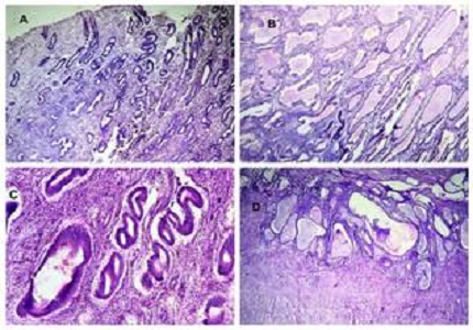
Results: Total 542 endometrial samples were received in department of Pathology during September 2013 to September 2015.Out of 542 endometrial samples, 131 are endometrial lesions. Among 131 endometrial lesions, 115 are non-neoplastic while 16 are neoplastic lesions. Most common age group for non-neoplastic lesions is 41-50 years while for neoplastic lesions it is about 51-60 years. Adenomyosis the most common (11.99%) non-neoplastic lesion while endometrioid adenocarcinoma (46.67%) ismost common neoplastic lesion.
Conclusion: In present study, non- neoplastic lesions were more common out of all endometrial lesions. Among neoplastic lesions malignant lesions were more common than benign lesions; histopathology is important for ensuring diagnosis and deciding management, particularly for malignant diseases.
Downloads
References
Robbins and Cotran. The Female Genital Tract, In: Pathologic Basis of Disease, 8th edition; USA, Saunders Elsevier publisher 2010; 1024.
Prat J. Female Reproductive System, In Ivan Damjanov & James Linder edited Anderson’s Pathology, Vol. 2, 10th edition; USA, Mosby publisher 2009: 2061.
Robbins and Cotran. The Female Genital Tract, Pathologic Basis of Disease, 8th edition; USA, Saunders Elsevier publication 2010; 1026.
Juan Rosai. Female Reproductive System. Rosai and Ackerman’s. Surgical Pathology. Vol.2, 10th edition; USA, Mosby publication 2009; 1479.
Onuma K, Dabbs DJ, Bhargava R. Mammaglobin expression in the female genital tract: immunohistochemical analysis in benign and neoplastic endocervix and endometrium. Int J Gynecol Pathol. 2008 Jul;27(3):418-25. doi: 10.1097/PGP.0b013e31815d05ec.
Montes M, Roberts D, Berkowitz RS, Genest DR. Prevalence and significance of implantation site trophoblastic atypia in hydatidiform moles and spontaneous abortions. Am J Clin Pathol. 1996 Apr;105(4):411-6.
Sobande AA, Eskander M, Archibong EI, Damole IO. Elective hysterectomy: A Clinicopathological review from Abha Catchment area of Saudi Arabia. WAJM 2005; 24; No.1; Jan-March 2005: Page: 31-35.
Gowari M, Mala G, Murthy S, Nayak V. Clinicopathological Study of Uterine Leiomyomas in Hysterectomy Specimens. Journal of Evolution of Medical & Dental Sciences; Vol. 2; Issue 46; Nov.2013:Page: 9002-9008.
Kartikeyan MT, Kumar R, Thomas E. Clinicopathological study of hysterectomy among rural patients in a tertiary care center. IOSR J Dent Med Sci ver IV Volume 14, Issue 5 Ver. IV (May. 2015), PP 25-27.
Zill-E-Huma, Arjumand. A clinicopathological review of elective hysterectomies in sir Gangaram hospital. Pakistan J Med Heal Sci [Internet] 2012; 6: 970- 2.
Tiwana KK, Nibhoria S, Monga T, Phutela R. Histopathological audit of 373 non-oncological hysterectomies in a teaching hospital. Patholog Res Int; Sept.2014; 46: 871- 5. http://dx.doi.org/10.1155/2014/468715.
Pessoa JN, Freitas AC, Guimaraes RA, Lima J, Dos Reis HL, Filho AC. Endometrial Assessment: When is it Necessary? J Clin Med Res. 2014 Feb;6(1):21-5. doi: 10.4021/jocmr1684w. Epub 2013 Dec 13.
Ates S, Ozcan P, Aydin S, Yardimci AS, Karaca N, Kilic G, Sevket O. Histopathologi¬cal analysis of 422 non-oncological hysterectomies in a university hospital. J Clin Anal Med 2015; DOI: 10.4328/ JCAM. 3505.
Surti HB, Zaveri P, Shah CK, Shah NR. Clinicopathological study of hysterectomy cases. Biennial Journal of GAPM 2015: 1- 4. Doi: http://dx.doi.org/10.18203/2320-1770.ijrcog20170575.
Bodal VK, Kaur N, Das T, Bal MS, Suri AK, Kaur S: Medical correlation of the various clinical findings & chief complains with histopathological pattern of endometrium biopsies: A study of 300 Cases. Research and reviews; Volume 3 | Issue (Supplement 3) | July - September, 2014: page no. 39- 45.
Wader JV, Jain A, Kumbhar SS, Vhawal V. Histiocytic endometritis. Am J Case Rep. 2013 Aug 26;14:329-32. doi: 10.12659/AJCR.889248. eCollection 2013.
Latha PS, Chaitanya B, Reddy S. Histopathological prognostic factor comparison of endometrial cancer patients in a tertiary hospital in India. Int J Reprod Contraception, Obstet Gynecol; Mar. 2014; 3 (1): 102-104. Doi: 10.5455/2320-1770.ijrcog20140320.
Deodhar KK, Rekhi B, Menon S, Ganesh B. An audit of histopathology reports of carcinoma endometrium: Experience from a tertiary referral center. J Postgrad Med; Apr-Jun 2015; 61(2): 84-87. Doi: https://dx.doi.org/10.4103%2F0022-3859.150444.
Himel B, Dewan. Clinicopathological study of endometrium in peri-menopausal and post-menopausal women in a tertiary care hospital in eastern India. IOSR J Dent Med Sci; Feb. 2014; Vol. 13 Issue 1 ver. X: 16- 23.

 OAI - Open Archives Initiative