E-ISSN:2456-1487
P-ISSN:2456-9887
RNI:MPENG/2017/70771

Research Article

Histopathological

Tropical Journal of Pathology and Microbiology

2020 Volume 6 Number 3 March
Publisherwww.medresearch.in

Histopathological study of ovarian lesions at a tertiary rural hospital

Gaikwad SL.1, Badlani KS.2*, Birare SD.3
DOI: https://doi.org/10.17511/jopm.2020.i03.06

1 Sheela Lakshmanrao Gaikwad, Associate Professor, Swami Ramanand Teerth Rural Government Medical College, Ambajogai, Maharashtra, India.

2* Koshu Sitaram Badlani, Post-graduate, Swami Ramanand Teerth Rural Government Medical College, Ambajogai, Maharashtra, India.

3 Shivaji Dadarao Birare, Professor, Swami Ramanand Teerth Rural Government Medical College, Ambajogai, Maharashtra, India.

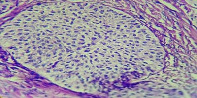
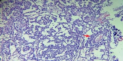
Background:Ovaries are the third leading site of cancer among women trailing behind cervical and breast cancer according to the Indian cancer registries. The spectrum of ovarian lesions is wide with harmless simple cystic lesions at one end and the fatal aggressive malignant lesions at the other end. Thus, ovarian neoplasm offers a good field for research. Methods: This was a prospective two years observational study and was conducted at the Department of Pathology SRTR GMC, Ambajogai, including 185 ovarian lesions. Result: Total 185 ovarian lesions were studied, 101 cases were non-neoplastic while 84 cases were neoplastic. The most common non-neoplastic lesion was corpus luteal cyst (27.7%) followed by simple ovarian cyst (24.7%) and follicular cyst (21.8%). Among 84 neoplastic cases, 74 cases (88.1%) were benign,02 cases (2.4%) were borderline tumours and 08 cases (9.5%) were diagnosed malignant. Conclusions: Both non-neoplastic as well as neoplastic lesions of ovary often present with similar clinical and radiological features. So histopathological study is essential to diagnose ovarian tumours and predict their prognosis.

Keywords: Ovarian Lesions, Non-Neoplastic, Neoplastic, Histopathological Study

Corresponding Author How to Cite this Article To Browse
Koshu Sitaram Badlani, Post-graduate, Swami Ramanand Teerth Rural Government Medical College, Ambajogai, Maharashtra, India.
Email:
Gaikwad SL, Badlani KS, Birare SD. Histopathological study of ovarian lesions at a tertiary rural hospital. Trop J Pathol Microbiol. 2020;6(3):245-252.
Available From
https://pathology.medresearch.in/index.php/jopm/article/view/441

Introduction

The ovary has three main histologic compartments i.e. the surface Mullerian epithelium, the germ cells and the sex-cord stromal cells. Each compartment gives rise to distinct non-neoplastic and neoplastic lesions [1,2]. Such lesions can be found from neonatal to postmenopausal ages and it accounts for around 30% of all female genital

cancers [3]. The incidence and prevalence of ovarian cancer vary in different geographical areas of the country. Indian trend analysis reveals a steady increase in the age-standardized incidence rate of ovarian cancer ranging from 0.26% to 2.44% per year in different area registries [4]. However, a few of the benign lesions can be confused with neoplasm clinically, intra-operatively or on histopathological examination.


Manuscript Received Review Round 1 Review Round 2 Review Round 3 Accepted
10-03-2020 20-03-2020 25-03-2020 29-03-2020
Conflict of Interest Funding Ethical Approval Plagiarism X-checker Note
No Nil Yes 15%

© 2020 by Sheela Lakshmanrao Gaikwad, Koshu Sitaram Badlani, Shivaji Dadarao Birare and Published by Siddharth Health Research and Social Welfare Society. This is an Open Access article licensed under a Creative Commons Attribution 4.0 International License https://creativecommons.org/licenses/by/4.0/ unported [CC BY 4.0].

Ovarian tumours include a complex, wide spectrum of neoplasms involving a variety of histological patterns ranging from epithelial tissues, connective tissues, specialized hormone-secreting germinal and embryonal cells [5]. The pathology of ovarian tumours and tumour-like conditions is one of the most complex areas in gynaecology. This is because the ovary produces a wide range and variety of tumours in the body relative to any other organ [6].In addition, the aetiology of ovarian cancers is poorly understood.Past epidemiological research focused on epithelial tumour aetiology and classified it as variables with increased risk of ovarian cancer, including a family history of ovarian cancer, advanced age, and nulliparity. Many factors known to be associated with decreased risk of ovarian cancer include a history of tubal ligation or hysterectomy, number of births, and the use of oral contraceptive (OC). There are several causes that have an inconclusive link to the incidence of ovarian cancers such as smoking, alcohol or coffee intake, fertility and infertility drug use, hormone replacement therapy (HRT), talc use, diet, obesity, first-born period, menarche/menopause period, breastfeeding [7-9].

The treatment and prognosis of ovarian neoplasms are based upon the accurate surgical staging and thorough pathological evaluation. Few organs exhibit as diverse a variety of tumour types as the ovary itself. For this cause, the surgical pathologist's differential diagnosis of ovarian tumours remains a daunting task.Nonetheless, significant progress has been made in the awareness of ovarian tumours and their molecular genetics and histopathological characteristics over the past few years and new therapeutic modalities have been developed accordingly [10]. The present study has been conducted with the aim to observe the frequency of neoplastic and non-neoplastic lesions in a tertiary care centre of rural India.

Material and Methods

Type of study and duration of study:After obtaining Institutional Ethical Committee approval, thisprospective observational study was carried out in a Department of Pathology at S.R.T.R. GMC Ambajogai, India during a period of two years from November 2017 to October 2019.

Study population: Total 434 ovarian specimens were received in the Department of Pathology during the study period but in 249 cases no specific

pathology other than normal findings was observed. Hence, only 185 cases were included in the study.

Inclusion criteria were all the ovarian specimens received in the Pathology department in the form of resected ovarian masses/ cystectomy specimens, ovarian biopsy specimen, tubo-ovarian masses and hysterectomy with salpingo-oophorectomy specimen.

Normal ovaries and specimens other than ovaries and ovarian specimens received after the study period were excluded from the study. Surgical specimens were obtained in fixative (10% formalin). Detailed clinical history, investigation, the radiologic finding was noted in each patient. Specimen were identified, specimen number was given to each specimen. Type of specimen was identified. Gross examination was done which included size, colour, and consistency and cut surface. The most representative areas of each specimen were found, and sections were taken of size 1.5x 1 cm. Tissue processing was done. The section was dehydrated in alcohol, cleared in xylol and embedded in paraffin wax. The corresponding blocks were prepared and 3 to 5-micron thick section were cut from each paraffin block and stained with hematoxylin and eosin stain [11]. Histopathological slides were examined by a senior pathologist;the correlation was done with clinical presentation. Data were entered into Microsoft Excel to calculate number and percentages.

Results

In the present study of 2 years from November 2017 to October 2019, in total 434 ovarian specimens were received. Out of 434 specimens, 249 showed no specific pathology, other than normal findings. Hence, only 185 cases were included in the present study, with 101 non-neoplastic ovarian lesions and 84 neoplastic ovarian lesions(Table 1).

Table-1: Distribution of Neoplastic and Non-Neoplastic Ovarian lesions.

Lesions No. of cases Percent
Non-Neoplastic lesions 101 54.6%
Neoplastic lesions 84 45.4%
· Benign 74 (88.1%) 40%
· Borderline 02 (2.4%) 1.1%
· Malignant 08 (9.5%) 4.3%
Total 185 100%

Table-2: Distribution of Ovarian Tumours according to histopathological type.

Nature of tumour No. of cases Percentage
I. Surface Epithelial Tumours 63 75%
A. Serous Tumours
· Serous Cystadenoma 30 35.7%
· Serous Borderline Tumour 01 1.2%
· Papillary Serous Carcinoma 02 2.4%
B. Mucinous Tumours
· Mucinous Cystadenoma 24 28.5%
· Mucinous Borderline Tumour 01 1.2%
· Mucinous Cystadenocarcinoma 02 2.4%
C. Transitional Tumours
· Benign BrennersTumour 02 2.4%
D. Endometrioid Carcinoma 01 1.2%
II. Sex Cord Stromal Tumours 04 4.8%
· Fibroma- Thecoma Group 04 4.8%
III. Germ Cell Tumours 17 20.2%
· Dysgerminoma 01 1.2%
· Yolk Sac Tumour 01 1.2%
· Mature Cystic Teratoma 14 16.6%
· Immature Teratoma 01 1.2%
Total 84 100%

In the present study, most common non-neoplastic lesion observed was corpus luteal cyst (28; 27.7 %) followed by simple ovarian cyst (25; 24.7%) and follicular cyst (22; 21.8%). The haemorrhagic cyst was observed in 19.8% cases, endometriosis in 4% cases and chronic oophoritis in 2% cases.

Out of 84 cases of ovarian tumours, surface epithelial tumours (63; 75%) were most common followed by germ cell tumour (17; 20.2%) and sex cord-stromal tumours (04; 4.8%)(Table 2).

In the present study, serous cystadenoma was the most common benign ovarian tumour contributing 40.5% cases, followed by mucinous cystadenoma (32.4%), mature cystic teratoma (18.9%), sex-cord stromal tumours (5.5%) and benign Brenner tumour (2.7%).

patho_441_01.jpg

Fig-1: Serous Borderline Tumour: Cut surface

shows papillary excrescences.

patho_441_02.jpg

Fig-2: Mucinous Cystadenocarcinoma: Cut surface showing multi-loculated cyst, the central solid area with areas of haemorrhage and necrosis.

patho_441_03.jpg

Fig-3: Immature Teratoma: The cut surface is partly solid and partly cystic. Multiple haemorrhagic cysts, hairs and whitish solid area can be appreciated.

Amongst surface epithelial tumour, serous cystadenoma constituted 47.6% cases followed by mucinous cystadenoma (38.1%).2 cases of borderline tumours were studied, one case of each serous borderline tumour and mucinous borderline tumour were observed.Only one case of benign Brenner tumour (1.6%) was noted. Amongst 5 cases of malignant surface epithelial tumours, serous carcinoma (2; 3.2%), mucinous adenocarcinoma (2; 3.2%) and endometrioid carcinoma (1; 1.6%) were studied.

The most common germ cell tumour observed in the present study is mature cystic teratoma (14; 16.6%). One case of each immature teratoma, dysgerminoma and yolk sac tumour was noted. All the 4 cases of sex-cord stromal tumours in the present study were fibro-thecoma.

Discussion

The ovary is the only organ of the body in which the tumour shows a wide histological variation, different behaviour and various histomorphological features.


Clinico-morphological features are helpful for the diagnosis and management of patients as they give an idea of disease progression. In the present study, a total of 4950 specimens were received, out of which 434 were specimens of the ovary, constituting 8.76% which is similar tostudy done by Parvatala et al [12].

Out of 434 specimens, 249 showed no specific pathology, other than normal findings. Hence, only185 ovarian lesions were included in the study of which 101 (54.6%) were non-neoplastic lesions and 84 (45.4%)were neoplastic ovarian lesions.This finding is in accordance with the previous studies [12-15].

In all these studies non-neoplastic lesions were more as compared to neoplastic lesions. Most of the non-neoplastic lesions were incidentally detected

(42.6%) with either hysterectomy or in pregnant patients during caesarean section or salpingo-oophorectomy in case of Ectopic pregnancy. Corpus luteal cysts were the most common non-neoplastic lesion in the current study (27.7%) followed by simple ovarian cysts (24.7%), this incidence iscomparable with the other studies [14-16] (Table-3). All the cases of corpus luteal cyst were less than 10 cms in diameter as similar tothe study done by Zaman et al [17].

Amongst 84 cases of ovarian neoplasms, 74 (88.1%) were benign, 2 (2.4%) were borderline and 8 cases (9.5%) were malignant lesions. Likewise, various studies showed that benign ovarian tumours were the most common ovarian neoplasms and malignant ovarian tumours were in the range of 4-21% and least common borderline tumours showed incidence ranging from 2-5% [15,18,19] (Table 4).


Table 3: Comparison of non-neoplastic ovarian lesions.

Lesions Present Study Hardik Makwana et al [14] Singh M et al [15] Sawant A and Mahajan S. [16]
Corpus Luteal Cyst 27.7% 29.95% 31.3% 12.7%
Simple Ovarian Cyst 24.7% 28.43% 18.2% 10%
Haemorrhagic Cyst 19.8% 17.77% 16.2% 5.5%
Follicular Cyst 21.8% 19.29% 34% 70%
Oophoritis 2% 2.03% - -
Endometriosis 4% 2.03% 0.3% 1.8%
Miscellaneous - 0.50% - -

Table-4: Comparison of benign, borderline and malignant ovarian neoplasms.

Studies Total No. of neoplastic lesions Benign(Percent) Borderline(Percent) Malignant(Percent)
Present study 84 88.1% 2.4% 9.5%
Singh M et al [15] 193 93.8% 2.1% 4.1%
Modepalli N and Venugopal SB [18] 141 83.01% 4.9% 12.1%
Sushama et al [19] 120 76.67% 2.50% 20.83%

Table-5: Comparison of Ovarian Tumours according to histopathological type.

Studies Surface epithelial tumours Germ cell Tumours Sex cord Stromal Tumours Metastatic Tumours
Present study 75% 20.2% 4.8% -
Kanthikar S N et al [13] 67.14% 22.85% 5.71% 4.28%
Sawant A and Mahajan S. [16] 84.8% 9.1% 6.1% -
Sushama et al [19] 70% 22.50% 5.83% 1.67%
Sudha V et al [20] 64.1% 26.1% 8.7% 1.1%


patho_441_04.jpg

Fig-4: Mature Cystic Teratoma: shows respiratory lining epithelium. Also shows sweat glands, hair follicles and adipocytes. (H and E: 10X).

patho_441_05.jpg

Fig-5: Benign BrennersTumours: shows islands of transitional epithelium. The individual tumour cells have abundant cytoplasm with nuclear grooves. (H and E: 40X).

patho_441_06.jpg

Fig-6: Yolk Sac Tumour shows the reticular arrangement of tumour cells with Schiller-Duval body (arrow). (H and E: 10X).

In the existing study, benign ovarian tumours were most commonly seen in the age group of 21-30 years and were more common unilaterally (70; 94.6%) than bilaterally (4; 5.4%).

Whereas malignant ovarian tumours were most commonly observed in the group of 51-60 years and 75% cases of malignant tumours were unilateral and 25% were bilateral.

According to histopathological type, the surface epithelial tumours (75%) were the most common type of ovarian neoplasm followed by germ cell tumour (20.2%) and sex cord-stromal tumours (4.8%). These findings are correlated well with the previous studies [13,16,19,20] (Table 5).

Serous cystadenoma was observed to be the most common benign ovarian tumour in the present study (40.5%) as well as in the studies done by Makwana et al [14] and Prakashet al [21].

The incidence of serious cystadenocarcinoma and mucinous cystadenocarcinoma were the same (25 %) which was the most common malignant ovarian tumour as reported by Jhaand Karki [22].

The histological distribution of surface epithelial-stromal tumours showed that serous cystadenoma (47.6%) and mucinous cystadenoma (38.1%) was the most common tumour whichis comparable with the other studies [14,19].

On histopathology, most common germ cell tumour was mature cystic teratoma (82.3%) which is similar to the findings of Sudha et al [20] study (87.5%).

Limitation of the study- There wasvariationobservedinthe resultsof other studies and present study due to the small sample size

of the present study. So, there is a need to study our experience with a larger population of patients.

Conclusion

Non-neoplastic ovarian lesions were more commonly seen in the present study. Among neoplastic lesions, benign tumours were more common than malignant and surface epithelial tumours were the most common histologic type. Ovarian malignancies act as "Silent Killer," as they occur in the advanced stage. Both non-neoplastic and neoplastic ovarian lesions often present with similar clinical and radiological characteristics. Histopathological research is therefore important for the diagnosis and prediction of ovarian tumours.

The present study suggested that patients receive medical advice late in rural health facilities because of the geographic location, deprivation and analphabetism. So, awareness among public and doctors, educating people, passive surveillance and community screening facility will be helpful in early


detection of the ovarian lesions and tumours.

Author’s contributions

Dr. Sheela L. Gaikwad:Analysis and preparation of the manuscript and critical revision.

Dr.Koshu S. Badlani: Data collection, analysis and preparation of the manuscript.

Dr. Shivaji D.Birare: Analysis of manuscript and critical revision.

Reference

  1. Prat J, D'Angelo E, Espinosa I. Ovarian carcinomas- at least five different diseases with distinct histological features and molecular genetics. Human Pathol. 2018;80;11-27.
  2. Supriya BR, Patel R, Patel M. Histopathological evaluation of Non-Neoplastic and Neoplastic Lesions of Uterine Cervix at tertiary care centre. Trop J Path Micro. 2019;5(3)177-182.
  3. Scully RE, Young RH, Clement PB. Tumors of the ovary, maldeveloped gonads, fallopian tube, and broad ligament. Amer Reg Pathol. 1998.
  4. Sharma I, Sarma U, Dutta UC. Pathology of ovarian tumour- A hospital-based study. Int J Med SciClin Invention. 2014;1;284-286.
  5. Selvaggi SM. Tumors of the ovary, maldeveloped gonads, fallopian tube, and broad ligament. Arch Pathol Lab Med. 2000;124(3)477.
  6. Murthy NS, Shalini S, Suman G, Pruthvish S, Mathew A. Changing trends in incidence of ovarian cancer – The Indian scenario. Asian Pac J Cancer Prev. 2009;10(6)1025-1030.
  7. Patel A, Patel NV. Study of frequency and histopathological pattern of soft tissue tumours in tertiary care centre of Gandhinagar, Gujarat. Trop J PatholMicrobiol. 2019;5(1)20-25.
  1. Kumar P, Malhotra N. Jeffcoate’s principles of gynaecology. Jaypee Brothers Medical Publishers (P) Ltd, 7th ed. 2008.
  2. Pradhan A, Sinha AK, Upreti D. Histopathological patterns of ovarian tumors at BPKIHS. Health Renaissance. 2012;10(2)87-97.
  3. Chae MS, Kim JW, Jung JY, Choi HJ, Chung HS, Park CS, et al. CT and MR imaging of ovarian tumours with emphasis on differential diagnosis. Radiograph. 2002;22(6)1305-1325.
  4. Ness RB, Grisso JA, Cottreau C, et al. Factors related to inflammation of the ovarian epithelium and risk of ovarian cancer. Epidemiol. 2000;11(2)111-117.
  5. Zhang J, Ugnat AM, Clarke K, Mao Y. Ovarian cancer histology specific incidence trends in Canada 1969-1993- age-period-cohort analyses. Br J Cancer. 1999;81(1)152-158.
  6. Ellenson LH, Pirog EC, Kumar V, Abbas AK, Fausto N, Aster JC (Eds). The female genital tract- Robbins and Cotran Pathologic Basis of Disease. 8th ed; Philadelphia, Elsevier Saunders. 2010;1039-1052.
  7. Prat J. Pathology of the ovary. Philadelphia- WB Saunders Company. 2004; p;223–7.
  8. Bancroft JD, Gamble M. Theory and practice of histological techniques. Elsevier Health Sci. 2008.
  9. Parvatala A, Prasad JR, Rao NB, Ghanta S. Study of Non-Neoplastic Lesions of the Ovary. IOSR J Dent Med Sci. 2015;14(1)92-96.
  10. Jha R, Karki S. Histological pattern of ovarian tumors and their age distribution. Nepal Med Coll J. 2008;10(2)81-85.

  1. Kanthikar SN, Dravid NV, Deore PN, Nikumbh DB, Suryawanshi KH. Clinico- histopathological analysis of neoplastic and non-neoplastic lesions of the ovary- a 3-year prospective study in Dhule, North Maharashtra, India. J Clin Diagn Res- JCDR. 2014;8(8)FC04–FC07.
  2. Makwana H, Maru A, Lakum N, Agnihotri A, Trivedi N, Joshi J. The relative frequency and histopathological pattern of ovarian masses – 11-year study at tertiary care centre. Int J Med Sci Public Health. 2014;3(1)81-84.
  3. Singh M, Jha KK, Kafle SU, Rana R, Gautam P. Histopathological Analysis of Neoplastic and Non-Neoplastic Lesions of Ovary- A 4 Year Study in Eastern Nepal. Birat J Health Sci. 2017;2(2)168-174.
  4. Sawant A, Mahajan S. Histopathological study of ovarian lesions at a tertiary health care institute. MVP J Med Sci. 2017;4(1)26-9.
  5. Zaman S, Majid S, Hussain M, Chughtai O, Mahboob J, Chughtai S. A retrospective study of ovarian tumours and tumour-like lesions. J Ayub Medical College Abbottabad. 2010;22(1)104-108.
  6. Mode Palli N, Venugopal SB. Clinicopathological study of surface Epithelial tumours of the Ovary- An institutional study. J Clin Diagnos Res- JCDR. 2016;10(10)01-04.
  7. Chandekar SA, Deshpande SA, Muley PS. A Clinico-Pathological Study of 120 Cases of Ovarian Tumors in a Tertiary Care Hospital. Int J Contemp Med Res. 2018;5(5)9-13.
  1. Prakash A, Chinthakindi S, Duraiswami R, Indira V. Histopathological study of ovarian lesions in a tertiary care center in Hyderabad, India-a retrospective five-year study. Int J Adv Med. 2017;4(3)745-749.
  2. Sudha V, Harikrishnan V, Sridevi M, Priya P. Clinicopathological correlation of ovarian tumors in a tertiary care hospital. Indian J Pathol Oncol. 2018;5(2)332-337.