E-ISSN:2456-1487
P-ISSN:2456-9887
RNI:MPENG/2017/70771

Case Report

Unveiling the Enigma

Tropical Journal of Pathology and Microbiology

2025 Volume 11 Number 1 Jan-Jun
Publisherwww.medresearch.in

Unveiling the Enigma: Case Series of Retroperitoneal Tumors and literature review

Das R1*, Purkayastha P2, Das R3
DOI:https://doi.org/10.17511/jopm.2025.i01.02

1* Rama Das, Department of Pathology, NBMC Darjeeling, West Bengal, India.

2 Pallabi Purkayastha, Department of Pathology, NBMC Darjeeling, West Bengal, India.

3 Rithuparna Das, Department of Pathology, NBMC Darjeeling, West Bengal, India.

Introduction: Retroperitoneal tumours (RPTs) are a diverse group of lesions that arise in the retroperitoneum and pelvic space. The histological spectrum of retroperitoneal masses is broad, ranging from major aggressive malignancies to benign lesions.

Objective: This case series and literature review aimed to study the incidence of retroperitoneal tumours presenting in a diverse array encountered in clinical practice, diagnosed on cytology or biopsy.

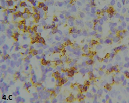
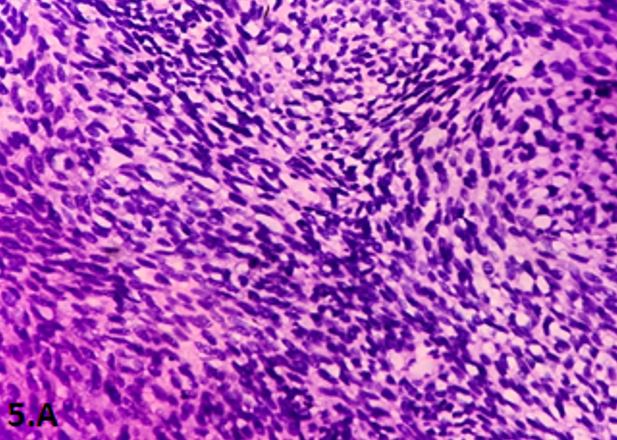
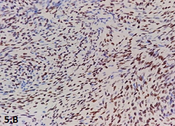
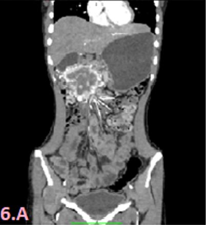
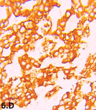
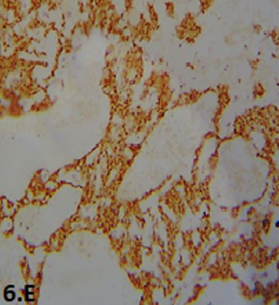
Materials and Methods: A retrospective observational study of 12 cases conducted over the last 2 (2023-2025) years. The RPTs diagnosed by guided Fine needle aspiration cytology or biopsy belonged to an age range of 7 years to 52 years. The clinical, imaging, cytomorphological and histopathological findings were studied. Immunohistochemical correlation was performed wherever available.

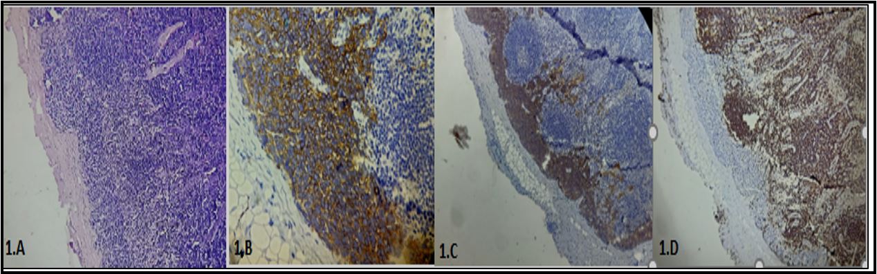
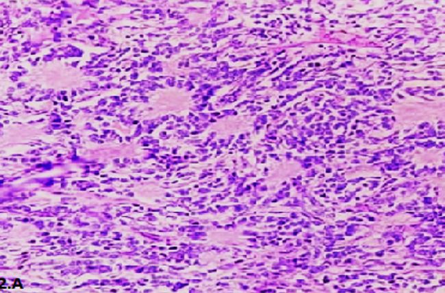
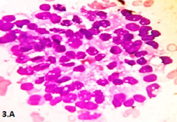
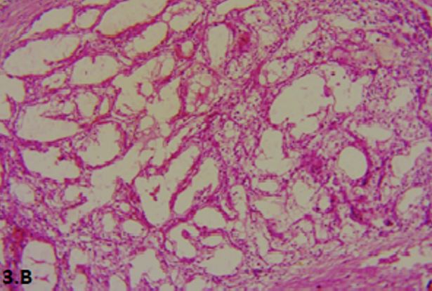
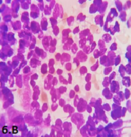
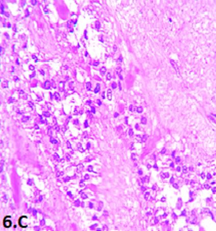
Results: We identified two cases of Neuroblastoma and two cases of Solid pseudopapillary pancreatic tumour with different presentations. One was a child and the other was an adolescent among neuroblastomas. Other patients presented with abdominal or left flank mass, left hypochondrial pain and were diagnosed as Paraganglioma, Multilocular Cystic Renal Neoplasm of Low Malignant Potential, Extraskeletal Ewing’s sarcoma, Synovial sarcoma, and Intraductal papillary mucinous neoplasm of pancreas. Among three cases of one unique case of Renal cell carcinoma was presented as a metastatic swelling in the left arm.

Conclusion: The cases presented in this series underscore the diverse nature of retroperitoneal masses, ranging from benign tumours to highly aggressive malignancies.

Keywords: Retroperitoneal tumours, Malignant, hypochondrial pain, metastatic arm swelling

Corresponding Author How to Cite this Article To Browse
Rama Das, , Department of Pathology, NBMC Darjeeling, , West Bengal, India.
Email:
Das R, Purkayastha P, Das R, Unveiling the Enigma: Case Series of Retroperitoneal Tumors and literature review. Trop J Pathol Microbiol. 2025;11(1):5-16.
Available From
https://pathology.medresearch.in/index.php/jopm/article/view/680

Manuscript Received Review Round 1 Review Round 2 Review Round 3 Accepted
2025-06-02 2025-06-09 2025-06-16 2025-06-23 2025-06-30
Conflict of Interest Funding Ethical Approval Plagiarism X-checker Note
None declared No financial support taken Not required in our institution for case series 12.36

© 2025 by Das R, Purkayastha P, Das R and Published by Siddharth Health Research and Social Welfare Society. This is an Open Access article licensed under a Creative Commons Attribution 4.0 International License https://creativecommons.org/licenses/by/4.0/ unported [CC BY 4.0].

Download PDFBack To ArticleIntroductionAims and objectivesMaterials and methodsResultsDiscussionConclusionsReferences

Introduction

Diagnosis of Retroperitoneal tumours (RPTs) is quite challenging for Pathologist. Retroperitoneal space extends from lumbar to iliac region behind perit-oneal covering & is restricted behind by posterior abdominal wall. Its contents are pancreas, adrenal glands, kidneys, ureters, aorta & its branches, inferior vena cava & numerous lymph nodes enmeshed in loose fibroconnective tissue. RPTs can originate from their content or supporting soft tissue. These tumours are rare, heterogeneous & diverse group of lesions, comprising both benign & malignant, although malignancy is prevalent. RPTs often pose significant trouble in diagnosis for radiologists & pathologists [1], [2]. Though Histopa-thology examination is gold standard technique [3], relevant ancillary tests like guided FNAC, serum markers, & immunohistochemistry also act as helping hands for accurate diagnosis. Here, authors will share their experiences, diagnostic challenges, and knowledge in case series of 12 cases of RPTs. Research articles published in literature have dealt mostly with retroperitoneal sarcomas, while publica-tions on RPT case series as whole are rare. Our study is first to be reported from Northern Districts of West Bengal, locally known as North Bengal.

Aims and objectives

The main objectives were to report the occurrence of RPTs offering challenges in clinical diagnosis, which were finally confirmed on biopsy and IHC.

Materials and methods

A Retrospective observational study was conducted for the last 2years (2023-2025) in the Department of Pathology in a tertiary care hospital of Darjeeling, comprising 12 cases. The clinical features, imaging findings, and patients' demographics were collected from record sections, and paraffin blocks were studied for histopathological examinations. Immunohistochemical (IHC) correlation was performed wherever necessary in this series.

Results

Neuroblastoma: 2 cases

A 7-year-old boy presented with a history of fever, weight loss, fatigue, and severe anaemia for the last six months.

His haemoglobin level was 7g%, though HPLC was reported as a normal haemoglobin variant. On clinical examination, a hard, non-tender lymph node was palpable in the left lower neck region. The lymph node biopsy was done, which showed partial subcapsular region effacement by small round blue cells [fig.1 A-D]. Those small, round blue tumour cells were strongly positive for synaptophysin and chromogranin and negative for CD45. Thus, the IHC profile proved the metastasis of neuroblastoma in the cervical lymph node in that child. The patient was advised to undergo urgent computed tomography (CT) of the whole abdomen, and a retroperitoneal mass was detected on the left side of the abdomen, superior to the kidney. The patient was subsequently planned for midline laparotomy. The resected tumour showed histopathological features of neuroblastoma primarily located in the right retroperitoneal region, as corroborated by IHC.

Another 15-year-old male patient presented with an abdominal lump in the left iliac fossa for the last two months associated with burning micturition. A CT scan of the abdomen showed a retroperitoneal SOL posterior to the urinary bladder. The patient had undergone an exploratory laparotomy. The HPE showed the presence of a small round cell tumour with pseudo-rosette formation [fig.2A]. The neoplastic cells were strongly positive for Synaptophysin and Chromogranin A. Thus, consistent with neuroblastoma.

Clear Cell Renal cell carcinoma: 3 cases

In our case series, we have encountered one unique case of RCC among three cases that presented primarily as a metastatic swelling in the left arm of a 50-year-old male patient. It was a painless, soft tissue nodule for the last 3months. Initially, FNAC was performed to rule out soft tissue lesions, but cytomorphology revealed tumour cells in clusters and aggregates with abundant, pale to vacuolated cytoplasm, indistinct cell borders, round, haphazard nuclei, and variable, prominent nucleoli, which raised suspicion [Fig. 3A]. The background shows vascular stromal components and necrohaemo-rrhagic areas. The patient was advised to undergo radiological imaging of the lower abdomen in search of the primary lesion. The excisional biopsy of that nodule showed compact nests and sheets of tumour cells with clear cytoplasm and distinct membranes, surrounding a network of arborising small, thin-walled vessels [Fig 3B].


These tumour cells are positive for PAX8 and CD10 on IHC [Fig 3C,3D]. Subsequently, a CT scan of the lower abdomen found a solid mass in the left kidney. The diagnosis of metastatic clear cell carcinoma of the kidney was primarily made. The other two cases of 58 and 60 years, Male patients presented with right lower abdominal pain and a right kidney lump. Radical nephrectomy was done in all three cases. HPE of the excised renal tumours showed histomorphology of clear cell RCC, as well as positive IHC markers, similar to the HPE features of the excised metastatic nodule in the first case.

Extra-skeletal Ewing’s sarcoma (EWS): 1 case

A 52-year-old female presented with abdominal pain & discomfort for 3 months. She had recent history of loss of weight. All laboratory tests, including pregnancy test, glucose, & tumour markers [carcin-oembryonic antigen (CEA), carbohydrate antigen (CA19-9), cancer antigen (CA125), & alpha fetopro-tein (AFP)] were normal. CT of abdomen & pelvis revealed mass (8x7 cm) in right pelvic retroperi-toneum region. Tru-cut biopsy revealed cytomor-phological diagnosis of small round blue cell tumour [Fig.4A] with possibility of extra-skeletal Ewing's sarcoma. Diagnosis was further substantiated by PAS & CD99 positivity [Fig.4B-C] & was negative for desmin and CK. FISH study for 22q12 (EWSR1) translocation was found to be positive.

Synovial Sarcoma (SS): 1 case

A 32-year-old male presented with persistent abdominal pain and palpable lump in right flank. His medical history was unremarkable. On physical examination, firm, non-tender lump was palpated in right upper quadrant. CT scan of abdomen revealed an irregular heterogeneous mass in right retroperitoneum measuring 13 cm x 10 cm x 7cm, with increased vascularity, focal necrosis, & displacing surrounding structures. Histopathology is characterised by monomorphic spindle-shaped cells arranged in fascicles [fig.5A] with focal staghorn blood vessels & scattered mast cells. Immunohistochemical staining was positive for TLE1, BCL2, S100 and negative for CD34 [fig.5B], confirming diagnosis of synovial sarcoma.

Solid Pseudo Papillary Neoplasm of Pancreas (SPPN-P): 2 cases

A 14-year-old girl and a 27-year-old female, the first one presented with a history of dyspepsia and intermittent epigastric pain since 6 months,

the other one had a history of epigastric and right upper quadrant pain, nausea and vomiting since 2months. The CT scan revealed a defined, solid-cystic mass on the pancreatic head in both cases. [Fig.6A]. USG guided FNAC was performed in both cases, which showed cellular smears having loose cohesive clusters of tumour cells in delicate papillary fronds. Individual cells are uniform with a moderate amount of cytoplasm, perinuclear clear vacuolation, and round to oval nuclei with granular chromatin [Fig.6B]. An exploratory laparotomy was performed in both cases. Grossly, they were well-defined encapsulated masses. On HPE, the tumour was heterogeneous, comprising solid and pseudo-papillary areas. The tumour cells have round to elongated, uniform nuclei and characteristic longitudinal grooves. Areas of hyalinization. Degeneration and cystic changes were present. IHC picked the diagnosis of solid pseudopapillary neoplasm of pancreas(SPPN-P) as the tumour cells are Vimentin Positive and have aberrant nuclear Beta-catenin expression.[Fig.6C-E]

Paraganglioma: 1 case

A 59-year-old man, a known hypertensive, presented with pain in the left lumbar region. CT imaging showed a spherical lesion with rim enhancement close to the pancreatic tail. USG guided FNAC showed a cellular bloody smear, comprising poorly cohesive clusters of tumour cells having abundant, pale cytoplasm with indistinct cell borders forming a web-like background with fine red granulation in some of the cells. There is marked anisokaryosis with speckled chromatin and intranuclear pseudo-inclusions. The patient was planned for surgery and a biopsy. HPE shows ribbons, nests of round to oval polygonal cells separated by fibrovascular stroma (zellballen) pattern and often forming a pseudo-rosette. [Fig.7A] |he tumour cells are strongly positive for S100, GATA3 and chromogranin [Fig.7B, C] and negative for CD99, CD117.

Multilocular Cystic Renal Neoplasm of Low Malignant Potential (MCRNLMP): 1 case

A 45-year-old male patient presented with left flank pain and hypertension. CT scan showed an ill-defined diffuse mass in the left kidney with heterogeneous post-contrast enhancement. Radical nephrectomy was performed. It was a tan yellow multiloculated cystic mass 10x8 x4 cm grossly involving the entire kidney and perinephric fat.


HPE of mass showed multiple cysts separated by thin fibrous septa, lined by neoplastic cells in a single layer with clear cytoplasm with low nuclear grade (ISUP/WHO gd1) [Fig.8A] The tumor was positive for PAX8, CD10, EMA, CKAE1/AE3[Fig.8B], Final diagnosis of MCRNLMP was considered.

Intraductal Papillary Mucinous Neoplasm of Pancreas (IPMN): 1 case

A 75-year-old female patient presented with pain in the left hypochondrium.

On CT scan, multiloculated grape-like cystic mass arising from head & body of pancreas extending inf-eriorly along transverse mesocolon was seen. Pancreatic-splenectomy with left hemicolectomy was done. HPE showed multiloculated mucin-filled cystic tumour having tall cuboidal mucinous epithelial lining with focal branching papillary projections [Fig. 9A]. Low-grade dysplasia noted. No ovarian-type stroma found. Tumour was positive for CK7 & MUC2 [Fig.9B] & negative for MUC1. Therefore, final diagnosis of intest. type IPMN was rendered. 

patho-680-01..JPG
Fig. 1A:
Infiltration of small round blue cell tumour in the subcapsular region of a cervical lymph node (H&E,20X). 1B: The tumour cells are strongly Chromogranin A positive. Fig. 1C: The tumour cells show synaptophysin positivity. The metastatic tumour cells of neuroblastoma are CD45-negative, and the rest of the lymphoid cells take up the CD45 stain.

patho-680-02..JPG
Fig 2A:
The tumour cells of neuroblastoma are composed of small round cells, having indistinct cell borders and salt paper chromatin and forming Homer-Wright pseudorosettes with eosinophilic neurophil in the centre. ( H&E,20X)

patho-680-03..JPG

patho-680-04..JPG

patho-680-05..JPG


patho-680-06..JPG
Fig. 3A:
FNA smear of arm swelling showing clusters of tumour cells with indistinct cell borders and abundant vacuolated cytoplasm. The nuclei of these cells show moderate atypia, irregular contour and prominent nucleoli. ( 40X Leishman stain). 3B: The tumour cells with clear cytoplasm are arranged in nests with delicate septa and blood vessels (20X H&E stain). 3C: The tumour cells of RCC show diffuse nuclear positivity for PAX8. 3D: RCC tumour cells are cytoplasmic positive for CD10 with apical accentuation.

patho-680-07..JPG

patho-680-08..JPG

patho-680-09..JPG
Fig. 4A:
The sheets of tumour cells are uniformly sized small round cells, slightly larger than small lymphocytes. Stippled chromatin with inconspicuous nucleoli, indistinct cytoplasmic membrane and clear to eosinophilic scanty cytoplasm noted on HPE (20X H&E stain) 4B: The small round cells contain a significant amount of cytoplasmic PAS-positive material 4C: CD99 was found to have strong and diffuse membranous expression in tumour cells-diagnostic of EWS.

patho-680-10..JPG

patho-680-11..JPG


Fig. 5A: The monophasic Synovial Sarcoma tumour has hypercellular fascicular architecture with little intervening stroma. The tumour cell population are monotonous, having spindled nuclei with overlapping features and scant amphophilic cytoplasm (20X H&E stain). 5B: Almost 80% of tumour cells are nuclear positive for the specific TLE-1 IHC marker.

patho-680-12..JPG

patho-680-13..JPG
Fig. 6A:
CT scan of the whole abdomen showed a well-circumscribed, encapsulated, heterogeneous solid cystic mass arising from the tail of the Pancreas.

6B: FNAC smear revealed loose cohesive clusters of bland uniform tumour cells. These cells have round to oval nuclei with a groove and finely granular chromatin, a moderate amount of cytoplasm, and perinuclear clear vacuolation suggestive of SPPN-P (Leishman stain,40X)

patho-680-14..JPG

patho-680-15..JPG


patho-680-16..JPG
Fig. 6C:
HPE of SPPN-P showed a solid area of uniform tumour cells with areas of hyalinization and degeneration. The individual tumour cells have round to elongated nuclei, finely textured chromatin with often longitudinal grooves (40X H&E stain). 6D: The tumour cells are Vimentin Positive. 6E: The SPPN-P tumour cells showed aberrant positive nuclear Beta-catenin expression.

patho-680-17..JPG

patho-680-18..JPG

patho-680-19..JPG
Fig. 7A:
Distinct clusters and nests comprising epithelioid population of round to oval tumour cells separated by thin fibrovascular septa found in Paraganglioma of Retroperitoneal region (20X H&E stain) 7 7 7 7 7B: Paraganglioma cells are strongly nuclear reactive for GATA3. Fig. 7C: Diffuse cytoplasmic positive staining of Chromogranin - present in Paraganglioma cells.


patho-680-20..JPG

patho-680-21..JPG

patho-680-22..JPG
Fig.8A:
Variable-sized cysts with low-grade clear cell lining of MCRNLMP (20X H&E stain) in retroperitoneum.

8B: The nuclei of clear cells of MCRNLMP are positive for PAX8. 8C: The membranous expression of CD10 in the tumour cells confirms renal origin.

patho-680-23..JPG

patho-680-24..JPG
Fig. 9A: Pancreatic cystic mass lined by tall cuboidal epithelium with basally located nuclei without significant pleomorphism- HPE of intestinal type IPMN (40X H&E stain). 9B: The lining epithelium is MUC2+

Discussion

In our case series, the age range was 7 years to 50 years. We could find many rare tumours in the retroperitoneum, comprising malignant (50%), tumour of low malignant potential (40%) and benign tumours (10%) over a period of 2 years. Most of the primary retroperitoneal tumours are reported as malignant, similar to findings in previous case studies and series. The retroperitoneum is a quite large space, though its contents are heterogeneous, but kidney tumours were the most common, accounting for these case series.


RCC was the commonest malignant tumour found in the retroperitoneum, similarly in previous studies by Telagavi KU et al. ( 3) and Das A et al. (4). Metastases of Renal Cell Carcinoma (RCC) to subcutaneous tissue are rare, but can occur. While RCC commonly metastasises to lungs, liver, bones, and brain, subcutaneous soft tissue is an uncommon site. These metastases may present as subcutaneous nodules, often years after the initial diagnosis and treatment of RCC. In our case series, we have encountered one unique case of RCC among three cases that presented primarily as a metastatic swelling in the left arm. Initially, FNAC was done to rule out a soft tissue lesion. But cytomorphology showed the features of RCC. HPE and IHC played a pivotal role in our final assessment, ultimately in these cases. Olivia Kozak et al. and Walton J et al. reported [5], [6] a similar case of metastatic RCC in the forearm. The cause of soft tissue metastatic cancer deposits in the absence of an identifiable primary tumour is unknown, and the recommended evaluation includes history and physical examination and a CT scan of the abdomen & pelvis, followed by further studies tailored to the individual.

Neuroblastoma is derived from neural crest cells, so the tumour can arise not only from the adrenal gland but also anywhere along the sympathetic chain. Among two cases of neuroblastoma, we encountered one rare case of metastatic deposit of neuroblastoma in the left cervical lymph node. That was a boy, 7 years old, presented with a history of fever, weight loss, and severe anaemia. Initially, he was suspected of hemoglobinopathy. Only physical examination could reveal one enlarged cervical lymph node. The identification of small round blue cells infiltration in the subcapsular region on HPE of the lymph node among the lymphoid cell population gave credit for the ultimate diagnosis of metastatic deposits and detection of primary neuroblastoma in the right retroperitoneum on subsequent radio-imaging. Further support by IHC was considered for the final diagnosis. We could find one case study with 16% head and neck metastasis of neuroblastoma in children [7] and one case report of a similar presentation by Mousa AH et al. [8] without any primary tumour. EWS in the retroperitoneal region is very rare and usually presents with vague pain in the peritoneum. Radio-imaging can often localise the tumour, but excision biopsy with HPE ultimately concludes diagnosis [9].

AlRashed R and Wu, SY et al. have case reports of extraskeletal Ewing’s sarcoma in retroperitoneal location. [10], [11] and got a definitive diagnosis on tissue biopsy. EWSR1 rearrangement was first identified in Ewing's sarcoma, but these translocations were also found in other soft tissue tumours like Myxoid liposarcoma [12], Extraskeletal myxoid chondrosarcoma [13] and Clear cell sarcoma [14] in small percentages. In this case, we found nests, sheets of small, round cells separated by a thick fibrous band. The small cells have round nuclei, stippled chromatin and very scanty cytoplasm. So, the histomorphology along with relevant immunohistochemical markers, i.e CD 99, FLI-1, are needed for retroperitoneal EWS diagnosis.

SS is very uncommon in the retroperitoneal region. It can grow larger in this RP region and remains asymptomatic unless it compresses the adjacent organ. Histologically, the tumour was monophasic in this case series and was challenging to diagnose. The tumour cells were positive for S100, BCL-2, and TLE-1, which enabled us to reach the diagnosis of poorly differentiated SS. Sarra Ben Rejeb et.al and Ansari Djafari A et.al could report the case of huge monophasic RPSS separately with molecular diagnosis. [15], [16] by the former author. We have no scope for molecular analysis for SS in our institution. We could find two cases of SPPN-P in female patients. SPPT is a rare tumour of the pancreas, accounting for only 1-2% of all pancreatic neoplasms, with a preponderance in young women. [17] In our case series, we found similar cases in a very young girl of 14 years and a woman of 27 years. Most of the data on SPPT was a case report or a small case series. WHO has classified it as a low-grade malignant neoplasm in 2010 [18], but few cases are reported in the literature with malignant metastatic potential [19] with high serum CA125 and tumour size >5cm. The pathogenesis is still unclear, but it can arise from pluripotent stem cells in the retroperitoneum and could be due to mutations in the β-catenin gene. In this case series, both tumours were < 5cm in maximum diameter, and no metastasis was found. Retroperitoneal Paragangliomas are very rare tumours, generally arising from the chromaffin cells of the sympathetic chain and the adrenal gland. They can arise from an extra adrenal site in 15% of the population and remain asymptomatic in 10 to 30 % cases [20].


The symptoms may vary from palpitations, tachycardia, and hypertension. Our case was hypertensive and had only complained of vague abdominal pain. The tumour was biochemical pseudo-silent as preoperative 24-hour urine metanephrines were normal and plasma Chromogranin A level was mildly raised. CT scan of the abdomen showed a spherical lesion with rim enhancement close to the pancreatic tail. FNAC raised the suspicion for round cell tumours with differential diagnosis of neuroendocrine tumour of the tail of pancreas, gastrointestinal stromal tumour (GIST), or paraganglioma. As described by Ramnani S et. al. from India for a case of symptomatic retroperitoneal paraganglioma and Imad Ghantous et.al from Beirut.LBN, complete surgical resection of the tumour and meticulous pre-operative preparation remain a pivotal modality of treatment [21], [22], so complete resection was done in our case with special care for handling intraoperative hypertensive crisis. The ultimate tissue diagnosis was done by HPE and finally confirmed by IHC profile.

MCRNLMP is one type of RCC that is diagnosed on imaging as a multiloculated cystic lesion with thin septation. Radiological diagnosis often puts the cases in a diagnostic dilemma. Schieda N et. al used Bosniak classification [23], which predicts the chances of malignancy and categorises MCRNLMP as IIF to IV. The present case was a heterogeneous lesion detected on non-contrast CT imaging, but found as a large multiloculated cystic lesion during grossing. The differential diagnosis was clear cell RCC with cystic changes((cysts are filled with hemorrhage, necrotic debris; keyboard like arrangement of nuclei with variable grade), cystic Clear cell Papillary RCC( predominantly papillary architectures formed by clear cells with low nuclear grade ) cystic nephroma (single layer cysts lining with various morphology; presence of ovarian/hyalinized/myxoid stroma), tubulocystic carcinoma( no clear cells; eosinophilic cytoplasm with high-grade nuclei and prominent nucleoli ) and renal cortical cyst( no clear cells lining in the wall of cyst). The ultimate diagnosis was achieved on histomorphology: cysts with a single layer of clear cells, bland nuclei, and absence of expansile growth. Confirmation was done by IHC. The tumour cells of MCNLMP were positive for PAX8, CA-IX, CK7, EMA, with focal positivity for CD10 and negative for AMACR, HMB45, CK 34βE12.

The majority of reported cases of MCRNLMP are of low pathologic stage, but extension to perinephric fat is a unique and unusual presentation in our case series, like previous reporting by Murshed KA et.al [24].

IPMN is quite common in the old age group > 65 years, with a common symptom of upper abdominal pain. It can arise from - main duct, branched duct, multicentric and of three types on histomorphology- Gastric, Intestinal and Pancreaticobiliary. Malignancy predisposition [25] is found more with main duct IPMN (60%) than mixed, followed by branched (12-30%). In our series, it was a cystic mass filled with mucin involving the head and body of the pancreas. Low-grade dysplasia was found on microscopy. Histomorphology was of intestinal type IPMN, ascertained as tumour cells were positive for MUC-2and negative for MUC-1. This case was unique as a case report of branched duct-intestinal type IPMN is rare.

Limitation: The compiled and studied retroperitoneal cases in this period were small in sample size, as other cases couldn't be presented here due to incompleteness of required data and untraceable patients. The molecular and cytogenetic studies are not available in our institute.

Conclusions

The exact diagnosis of RPT is troublesome and quite challenging. Most of the RPTs are malignant, but as the space is large enough to allow the tumour growth silently, the clinical symptoms develop later. Guided FNAC often helps for initial diagnosis, but ultimately, HPE on biopsy is still considered the gold standard for diagnosis and aid for other ancillary investigations like IHC and molecular studies.

Funding: No financial support taken

Conflict of interest: None declared

Ethical approval: Not required in our institution for case series

References

1. Mota MMDS, Bezerra ROF, Garcia MRT. Practical approach to primary retroperitoneal masses in adults. Radiol Bras. 2018 Nov 23;51(6):391–400. [Crossref][PubMed][Google Scholar]


2. V Renuka I, Vaishnavi R, Sankar Naik B, Premalatha P, Ramya C. Rare tumours of retroperitoneum- A diagnostic challenge. IJPO. 2019 Nov28;6(4):671–6. [Crossref][PubMed][Google Scholar]

3. Telagavi KU, Bhavikatti BM, Reddy P. A Nine-Year Histopathological Retrospective Study of Retroperitoneal Masses. J Clin of Diagn Res. . 2022; 16(7): EC08EC12 [Crossref][PubMed][Google Scholar]

4. Das A, Gahine R, Patre V, Hussain N. Retroperitoneal tumour: A silent trespasser role of image-guided fine-needle aspiration cytology with histopathological correlation in early diagnosis. Acta Cytologica. 2019;63(3):189-97. [Crossref][PubMed][Google Scholar]

5. Kozak O, Turzyński P, Markiet K, Pieńkowska J, Skrobisz-Balandowska K, Studniarek M. Uncharacteristic metastasis of a renal clear-cell carcinoma to the muscles of the forearm—case report. BJR|case reports. 2017Sep;3(3):20150495. [Crossref][PubMed][Google Scholar]

6. Walton J, Li J, Clifton MM, Mori RL, Park AM, Sumfest JM. Metastatic clear cell renal cell carcinoma to the forearm without an identifiable primary renal mass. Urol Case Rep. 2019 Aug 6;27:100989. Doi: 10.1016/j.eucr.2019.100989. PMID: 31440453; PMCID: PMC6699464 [Crossref][PubMed][Google Scholar]

7. S Alvi, O Karadaghy, M Manalang, R. Weatherly Clinical manifestations of neuroblastoma with head and neck involvement in children Int J Pediatr Otorhinolaryngol, 97 (2017), pp. 157-162, 10. 1016/j. ijporl.2017.04.013 [Crossref][PubMed][Google Scholar]

8. Mousa AH, Baothman A, Alsayed AW, Rozan A, Ghannam YJ, Ibrahim A, Mostafa AF. Metastatic neuroblastoma presenting without a primary tumour: A case report. Radiol Case Rep. 2022 Apr 13;17(6):2137-2141. doi: 10.1016/j.radcr.2022.03.083. PMID: 35464796; PMCID: PMC9024373 [Crossref][PubMed][Google Scholar]

9. Choi J. H. , Ro J. Y. Retroperitoneal sarcomas: an update on the diagnostic pathology approach. Diagnostics. 2020 Sep;10(9):642. doi: 10.3390/diagnostics10090642 [Crossref][PubMed][Google Scholar]

10. AlRashed R, AlHarbi H, Abdulfattah F, Alhasan I, Alsannaa F. A case report of retroperitoneal Ewing sarcoma requiring adrenalectomy. Int J Surg Case Rep. 2022 Jun;95:106966. doi: 10.1016/j.ijscr.2022.106966. Epub 2022 Apr 11 [Crossref][PubMed][Google Scholar]

11. Wu, SY. , Hsu, CK. , Yue, CT. et al. Large retroperitoneal extraskeletal Ewing’s sarcoma with renal pedicle invasion: a case report. BMC Urol 23, 95 (2023) [Crossref][PubMed][Google Scholar]

12. Suzuki K, Matsui Y, Higashimoto M, Kawaguchi Y, Seki S, Motomura H, Hori T, Yahara Y, Kanamori M, Kimura T. Myxoid liposarcoma-associated EWSR1-DDIT3 selectively represses osteoblastic and chondrocytic transcription in multipotent mesenchymal cells. PLoS One. 2012;7(5):e36682. doi: 10.1371/journal.pone.0036682. Epub 2012 May 3. PMID: 22570737; PMCID: PMC3343026 [Crossref][PubMed][Google Scholar]

13. Hu HJ, Liao MY, Xu LY. Primary retroperitoneal extraskeletal mesenchymal chondrosarcoma involving the vena cava: A case report. Oncol Lett. 2014 Jun;7(6):1970-1974. doi: 10.3892/ol.2014.2012. Epub 2014 Mar 28. PMID: 24932271; PMCID: PMC4049765 [Crossref][PubMed][Google Scholar]

14. Katabuchi H, Honda R, Tajima T, Ohtake H, Kageshita T, Ono T, Okamura H. Clear cell sarcoma arising in the retroperitoneum. Int J Gynecol Cancer. 2002 Jan-Feb;12(1):124-7. doi: 10.1046/j.1525-1438.2002.01052.x. PMID: 11860547 [Crossref][PubMed][Google Scholar]

15. Sarra Ben Rejeb, Safia Sakly, Majdi Ben Romdhane, Mehdi Charfi, Adnen Chouchen. Primary retroperitoneal synovial sarcoma (RSS): A case report. International Journal of Surgery Case Reports. Volume 127,2025,110982, ISSN 2210-2612. [Crossref][PubMed][Google Scholar]

16. Ansari Djafari A, Razzaghi M, Rakhshan A, Faraji S, Rahavian AH, Hojjati SA. A Large Primary Retroperitoneal Synovial Sarcoma: A Case Report of a Huge Malignant Tumour. Iran J Med Sci. 2022 May;47(3):280-284. doi: 10.30476/IJMS.2021.90470.2141. PMID: 35634528; PMCID: PMC9126900 [Crossref][PubMed][Google Scholar]


17. Aktaş H, Işık Ö, Emiroğlu R. Solid pseudopapillary tumour of the pancreas: A case report. Turk J Surg. 2020 Mar 18;36(1):110-112. doi: 10.5578/turkjsurg.. 3237. PMID: 32637883; PMCID: PMC7315450 [Crossref][PubMed][Google Scholar]

18. Paluri RK, John TA, Anastasopoulou C, et al. Solid Pseudopapillary Epithelial Neoplasm (SPEN) of the Pancreas. [Updated 2024 Jan 10]. In: StatPearls [Internet]. Treasure Island (FL): StatPearls Publishing; 2025 Jan- [Crossref][PubMed][Google Scholar]

19. Chen L, Yuan M, Wang M, Luo C, Gao M, Huang Q, Li Z, Zhou Z. Case report: Retroperitoneal solid pseudopapillary neoplasm associated with multiple hepatic metastases. Front Oncol. 2024 Jan 30;14:1335930. Doi: 10.3389/fonc.2024.1335930. PMID: 38352895; PMCID: PMC10861742 [Crossref][PubMed][Google Scholar]

20. Rey Chaves CE, Ayala D, García G, Conde Monroy D, Sabogal Olarte JC. Retroperitoneal pheochromocytoma: Unusual presentation and atypical location. Int J Surg Case Rep. 2021 Aug;85:106248. doi: 10.1016/j.ijscr.2021.106248. Epub 2021 Jul 27. PMID: 34352621; PMCID: [Crossref][PubMed][Google Scholar]

21. Ramnani S, Chawla S, Utaal MS, Singh M, Adlakha D, Thakor M. A rare case of extra-adrenal retroperitoneal paraganglioma: case report. Int Surg J2024;11:644-8. . [Crossref][PubMed][Google Scholar]

22. Ghantous I, Najjar A, Tlaiss Y, Danaf S, Chouairy CJ. Resection of Retroperitoneal Extra-Adrenal Pheochromocytoma: A Case Report. Cureus. 2023 Aug 9;15(8):e43185. doi: 10.7759/cureus.. 43185. PMID: 37692674; PMCID: PMC10485873 [Crossref][PubMed][Google Scholar]

23. Schieda N, Davenport MS, Krishna S, et al. Bosniak classification of cystic renal masses, version 2019: a pictorial guide to clinical use. Radiographics 2021;41:814–828. . [Crossref][PubMed][Google Scholar]

24. Murshed KA, Akhtar M, Ben-Gashir M. Multilocular cystic renal neoplasm of low malignant potential extending into the perinephric adipose tissue. Hum. Pathol. : Case Rep. 019;17:200305 [Crossref][PubMed][Google Scholar]

25. Jadhav SP, Ali F, Kadam S, Chopra AK, Iyer VS. Intraductal papillary mucinous neoplasm in a 26-year-old lady. a diagnostic therapeutic challenge! Int Surg J 2022;9:746-8. . [Crossref][PubMed][Google Scholar]

Disclaimer / Publisher's Note The statements, opinions and data contained in all publications are solely those of the individual author(s) and contributor(s) and not of Journals and/or the editor(s). Journals and/or the editor(s) disclaim responsibility for any injury to people or property resulting from any ideas, methods, instructions or products referred to in the content.