E-ISSN:2456-1487
P-ISSN:2456-9887
RNI:MPENG/2017/70771

Case Report

Hematolymphoid Malignancies

Tropical Journal of Pathology and Microbiology

2024 Volume 10 Number 1 January-March
Publisherwww.medresearch.in

Hematolymphoid Malignancies with Neurological Manifestations and Hand-mirror cells in Peripheral Blood: A Report of two cases

Seshagiri K S1, Nandeesh BN2*, Devi B I3, Mallithavana S4
DOI:https://doi.org/10.17511/jopm.2024.i01.01

1 Sangeetha Seshagiri K, MD (Pathology), DCP, Clinical Pathologist, Specialist Grade, Department of Transfusion Medicine and Hematology, NIMHANS, Bengaluru, Karnataka, India.

2* B N Nandeesh, MD, DNB, PDF (Neuropathology), Additional Professor, Department of Neuropathology, NIMHANS, Bengaluru, Karnataka, India.

3 Indira Devi B, Mch Professor, Department of Neurosurgery, NIMHANS, Bengaluru, Karnataka, India.

4 S Mallithavana, DCP, Clinical Pathologist, Specialist Grade, Department of Transfusion Medicine and Hematology, NIMHANS, Bengaluru, Karnataka, India.

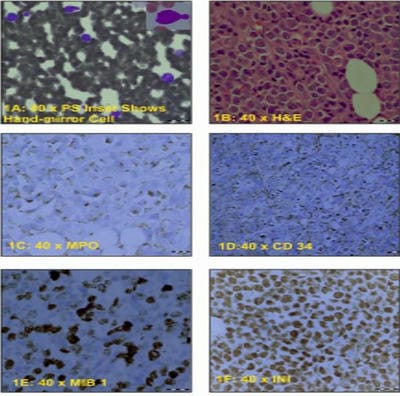
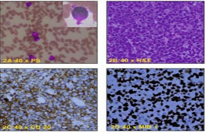
Hematolymphoid malignancies (HLM) can present with clinical features of systemic involvement and show hand-mirror cells (HMCs) in peripheral blood. Two patients presented with back pain and lower-limb weakness, blood smears revealed atypical cells, HMCs and thrombocytopenia. Imaging studies showed extradural spinal lesions. Histopathology of laminectomy specimens revealed myeloid sarcoma and diffuse B-cell lymphoma. HMCs can be seen in malignant and non-malignant conditions. The presence of HMCs may be the initial finding in peripheral blood indicating underlying malignancy with or without generalized symptomatology and leucocytosis. This study aims to emphasize that the presence of HMCs should prompt appropriate investigations and management after ruling out reactive conditions.

Keywords: Hematolymphoid Malignancies, Neurological Manifestations, Hand-Mirror Cells

Corresponding Author How to Cite this Article To Browse
B N Nandeesh, MD, DNB, PDF (Neuropathology), Additional Professor, Department of Neuropathology, NIMHANS, Bengaluru, Karnataka, India.
Email:
Seshagiri K S, Nandeesh BN, Devi B I, Mallithavana S, Hematolymphoid Malignancies with Neurological Manifestations and Hand-mirror cells in Peripheral Blood: A Report of two cases. Trop J Pathol Microbiol. 2024;10(1):1-4.
Available From
https://pathology.medresearch.in/index.php/jopm/article/view/641

Manuscript Received Review Round 1 Review Round 2 Review Round 3 Accepted
2024-02-01 2024-02-03 2024-02-10 2024-02-17 2024-02-24
Conflict of Interest Funding Ethical Approval Plagiarism X-checker Note
Nil Nil Yes 12.54

© 2024by Seshagiri K S, Nandeesh BN, Devi B I, Mallithavana Sand Published by Siddharth Health Research and Social Welfare Society. This is an Open Access article licensed under a Creative Commons Attribution 4.0 International License https://creativecommons.org/licenses/by/4.0/ unported [CC BY 4.0].

Introduction

Hematolymphoid Malignancies (HLM) involve myeloid and lymphoid cell lineages and affect blood, bone marrow, lymph nodes and lymphatic system. Clinically, they may present with features suggestive of involvement of various systems. Involvement of central nervous system (CNS) is not uncommon. These malignancies may present with hand-mirror cells (HMCs) in peripheral blood which can be the initial findings with or without other features of malignancies. This report aims to emphasize that the presence of HMCs should prompt the pathologist to guide the patients/clinicians to get appropriate investigations, workup and management.

Case report

Case 1: A 13-year-old male patient presented with back pain, tingling sensation, and rapidly evolving weakness in bilateral lower limbs. MRI showed multiple intracranial dural lesions and extradural spinal lesions. On examination, restricted eye movements, bifacial and neck flexor weakness were present. The patient had Haemoglobin (Hb) 10.1 gm/dl, Leukocyte count 10.0x10³ /µl, Platelet count 60x103 /µl. Peripheral smear revealed normocytic normochromic RBCs, normal leucocyte count with atypical mononuclear cells having increased nucleocytoplasmic ratio, nuclei with open chromatin, cytoplasm with few pink granules, several of them showing cytoplasmic projection from one pole of the cell – HMC, and thrombocytopenia (Figure:1A). Diagnosis of HLM was made and flow cytometry was considered. Flow-cytometric immunophenotyping showed a population of progenitors that expressed CD34, CD38, CD123, CD13, CD33, CD117, HLADR, cytoplasmic MPO showed aberrant expression of CD19, CD56 and cytoplasmic CD79a (subset). The immunophenotypic and clinical features suggested a diagnosis of acute myeloid leukaemia (AML) with t(8;21).

The patient underwent D6-D8 and D11-D12 laminectomy, and histopathologic examination revealed highly cellular hematopoietic neoplasm composed of sheets of cells infiltrating the fibroadipose tissue. The cells were large (2-3 times the size of RBCs) showing indented vesicular nuclei with prominent 2-3 nucleoli and abundant amphophilic to eosinophilic cytoplasm.

The brisk mitotic and apoptotic activity was seen with areas of karryorhectic debris (Figure:1B). Immunohistochemistry was positive for MPO and CD34 suggestive of myeloid neoplasm (Figures:1C&1D), and CD20, CD3, CD30, ALK were negative, ruling out a lymphoid neoplasm. MIB-1 labelling showed a high proliferative index which ranged from 55-60% (Figure:1E), and INI1 revealed retained expressionruling out Atypical Teratoid/Rhabdoid Tumor (Figure:1F). A diagnosis of myeloid sarcoma was considered.

patho_641_01.JPG
Figure:01 1A: Peripheral smear (PS) shows Normocytic Normochromic RBCs with atypical leucocytes showing high N:C. Inset shows a Hand mirror cells with cytoplasm at one pole. 1B: Histopathologic sections of laminectomy specimens revealed a highly cellular hematopoietic neoplasm composed of sheets of cells. The cells were large (2-3 times the size of RBCs) and showed indented vesicular nuclei with prominent 2-3 nuleoli and abundant amphophilic to eosinophilic cytoplasm. 1C & 1D: Immunohistochemistry (IHC) was positive for MPO and CD34 suggestive of myeloid neoplasm. 1E: MIB-1 labelling revealed high proliferative index. Features were suggestive of Myeloid sarcoma. 1F: INI1 showed retained expression (ruling out Atypical Teratoid/ Rhabdoid tumor)

Case 2: A 40-year-old female presented with bilateral lower limb weakness and pain in the lower back. MRI revealed a lesion at the D2/D3 vertebral region. Her haematological tests revealed Hb 11.5 gm/dl, Leucocyte counts 9.0x10³ /µl, Platelet count 56x103 /µl, peripheral smear


showing normocytic normochromic RBCs along with few normoblasts and leucocytes revealing predominantly increased mononuclear cells with a high nucleocytoplasmic ratio, open chromatin, prominent nucleoli, scant cytoplasm and coarse cytoplasmic granules, several of them having a cytoplasmic projection – HMCs and thrombocytopenia (Figure:2A). Diagnosis of hematolymphoid malignancy was considered. The D2/D3 laminectomy tissue on histopathological examination revealed an infiltrating tumour with high cellularity, composed of densely packed sheets of atypical cells with a lymphoid morphology. These cells showed scant cytoplasm and prominent nucleoli. Mitosis and apoptosis were brisk along with a sprinkling of reactive lymphocytes amongst atypical lymphocytes (Figure:2B). IHC revealed neoplastic cells which were diffusely positive for CD20 (Figure:2C). Also, reactive T cells were positive for CD3. CD138, synaptophysin and CK were negative. CD99 showed variable positivity. MIB-1 labelling showed a high proliferative index which ranged from 70-75% (Figure: 2D). The histology suggested a diagnosis of diffuse B cell lymphoma. The patient was discharged before flow cytometry was carried out and was lost for follow-up.

patho_641_02.JPG
Figure:02: 2A: PS showed Normocytic and Normochromic RBCs, atypical lymphocytes and thrombocytopenia. Inset shows a Hand mirror cells. 2B: Histopathological examination of laminectomy tissue revealed and infiltrating tumor with high cellularity, composed of densely packed sheets of atypical cells with lymphoid morphology. These cells showed scant cytoplasm and prominent nucleoli. Mitosis and apoptosis were brisk. 2C: IHC revealed neoplastic cells which were diffusely positive for CD20. 2D: MIB1 labelling showed high proliferative index. The histology was suggestive of Diffuse B Cell Lymphoma.

Prothrombin Time, Activated Partial Thromboplastin Time and Biochemical parameters were within normal limits in both cases.

Discussion

Leukemia can manifest with general and systemic clinical features that include involvement of bone marrow causing its failure, leading to fatigue and palpitations due to anemia, infections and fever due to neutrophilic dysfunction, bleeding due to thrombocytopenia and weight loss, anorexia and unexplained pain due to metabolic abnormalities. Leukemia can involve various systems. Leukemia involving CNS can present with features of raised intracranial tension like headache, altered mental status and sensorium, nausea, vomiting, and visual and hearing loss. Features of spinal cord compression like back pain, paraesthesia, limb weakness, and bowel and bladder disturbances may be encountered.[1] It is well known that leukemia has a high leucocyte count. However, few of them may present with normal leucocyte count.[2]

Systemic lymphomas and lymphatic leukaemia can cause intracranial, spinal epidural and peripheral nerve lesions causing back pain, lower limb weakness, and bladder disturbance etc., Bone marrow involvement can be present with secondary CNS lesions and epidural lymphomatous lesions. Further, NHL can cause haematological dissemination.[3] Lymphomas may primarily or secondarily involve the bone marrow and present with type B symptoms like fever, night sweats and weight loss with normal leucocyte count and Hb.[4] A significant number of lymphomatous cells may be seen in the leukemic phase.[5]

Pandey S et al, reported lymphomas, plasma cell neoplasms, and myeloid sarcomas presenting with epidural spinal lesions.[6] Both of our cases also presented with back pain and lower limb weakness with spinal lesions and normal leucocyte count. AML, CML, ALL, B-cell and T-cell NHLs can present with HMCs in peripheral blood showing round to oval nuclei with cytoplasmic projection from one pole.[7,8,9] However, HMCs are reported even in non-malignant conditions like Infectious mononucleosis.[10] Both of our cases showed HMCs in peripheral blood.

While evaluating the cases of HMC associated with malignancy, thorough clinical examination along with examination for necessary parameters that


include Auer rods, phi-bodies, N/C ratio, opened-up chromatin, nucleolus, clefted nuclei, WBC and platelet count, flow-cytometry and genetic studies have to be considered and correlated.

Conclusion

Hand mirror cells can be seen in both malignant and non-malignant conditions. Their presence may be the initial findings in peripheral blood with or without leucocytosis and type-B symptoms. Their presence should act as a clue for careful evaluation and correlation with the haematological and other investigations. If reactive conditions are ruled out, this finding may serve as a hint to consider evaluation of HLM, when clinical features and investigation findings favour. This article attempts to create awareness and indicate that morphological features/clinical signs act as important clues for some of the pathological conditions, including a malignant type.

References

1. Central nervous system involvement in adult acute lymphoblastic leukemia: diagnostic tools, prophylaxis, and therapy. Mediterr J Hematol Infect Dis. 2014; 6(1):e2014075. . [Crossref][PubMed][Google Scholar]

2. Acute leukemia presenting as bone pain with normal white blood cell count. Acute Med Surg. 2014;1(4):249. . [Crossref][PubMed][Google Scholar]

3. Involvement of the central nervous system in malignant lymphomas. Virchows Arch A Pathol Anat Histol. 1976;370(4):345-62. . [Crossref][PubMed][Google Scholar]

4. Clinical presentation and characteristics of lymphoma in the head and neck region. Head Face Med. 2019;15(1):1. . [Crossref][PubMed][Google Scholar]

5. Malignant Lymphoma: A Clinico-Pathologic Survey of 618 Cases. Am J Pathol. 1942:18(3):381-429. . [Crossref][PubMed][Google Scholar]

6. Hematolymphoid Malignancies Presenting with Spinal Epidural Mass and Spinal Cord Compression: A Case Series with Rare Entities. Ann Clin Lab Sci. 2019;49(6):818-828. . [Crossref][PubMed][Google Scholar]

7. Hand-mirror variant of microgranular acute promyelocytic leukemia. Leukemia. 1991;5(3):266-9. . [Crossref][PubMed][Google Scholar]

8. Hand-mirror-cell variant of B-cell lymphoma with hypercalcemia. Arch Intern Med. 1986;146(11):2273-4. . [Crossref][PubMed][Google Scholar]

9. Hand-mirror cell morphology of T-cell lymphoproliferative disorder presenting in pleural effusion with mediastinal widening. J Cancer Res Ther. 2011;7(3):389-90. . [Crossref][PubMed][Google Scholar]

10. Hand-mirror lymphocytes in infectious mononucleosis. Blood. 1980;55(6):925-30. . [Crossref][PubMed][Google Scholar]